

doi: 10.1101/gr.151700.
Construction and transposition of a 100-kilobase extended P element in Drosophila
Affiliations
- PMID: 11042158
- PMCID: PMC310958
- DOI: 10.1101/gr.151700
Item in Clipboard
Construction and transposition of a 100-kilobase extended P element in Drosophila
Genome Res.
2000 Oct.
Abstract
We have used P element deletion derivatives at defined locations in the Drosophila genome to construct a 100-kb extended P element more than twice the size of any previously available. We demonstrate that this prototypical extended P element is capable of transposition to new sites in the genome. The structural and functional integrity of a transposed extended P element was confirmed using molecular, genetic, and cytogenetic criteria. This is the first method shown to be capable of producing large, unlinked transpositional duplications in Drosophila. The ability to produce functional transposable elements from half-elements is novel and has many potential applications for the functional analysis of complex genomes.
Figures
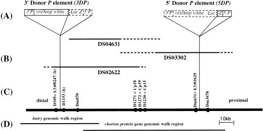
The genomic region encompassed by the extended P element. (A) The structure of the two P elements used to construct the extended P element is shown. The two closely linked P elements (3DP and 5DP) used for the construction of the extended P element contain the bacterial orgin of replication (ori), the bacterial ampicillin resistance gene (amp) and LacZ gene, and the Drosophila white gene, as well as the 5′ and 3′P element ends 3′P and 5′P (Haenlin et al. 1985). Dashed lines outline the regions of the P elements deleted during the production of half-P-element derivatives. (B) Position of P1 genomic phage in the 66D region used in this study. Uncertainty in the endpoints of P1 genomic clones are shown in dashed lines. (C) Genomic DNA in the 66D region (shown as a solid line) including chromosomal orientation (distal and proximal to the centromere) and the location of sequence tagged sites (sts) in the region, shown as solid circles. (D) Extent of two overlapping genomic λ phage walks through the region, used to characterize the extended P element further, are shown (Spradline 1981; Ish-Horowitz et al. 1985).
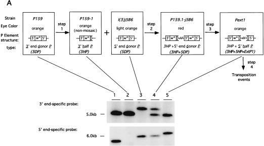
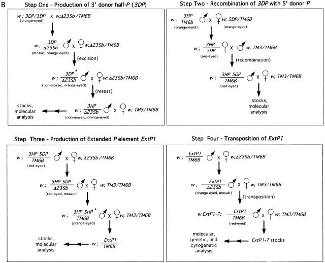
Construction and transposition of the prototypical extended P element, ExtP1. (A) Shown on the top are the three steps used to construct ExtP1. Each rectangular box shows the name of the strains utilized for each step followed by the eye color observed, structure of the P element, and type of P element. Southern blot analysis of the structure of each element, determined using 3′- and 5′-end-specific probes, is shown below. (B) Representative crosses carried out to perform each step shown in A. Abbreviations for balancer chromosomes used are: TM3 = TM3, SB ry e and TM6B = TM6B, Tb Hu e. All other abbreviations are given in A and in Results.
Construction and transposition of the prototypical extended P element, ExtP1. (A) Shown on the top are the three steps used to construct ExtP1. Each rectangular box shows the name of the strains utilized for each step followed by the eye color observed, structure of the P element, and type of P element. Southern blot analysis of the structure of each element, determined using 3′- and 5′-end-specific probes, is shown below. (B) Representative crosses carried out to perform each step shown in A. Abbreviations for balancer chromosomes used are: TM3 = TM3, SB ry e and TM6B = TM6B, Tb Hu e. All other abbreviations are given in A and in Results.
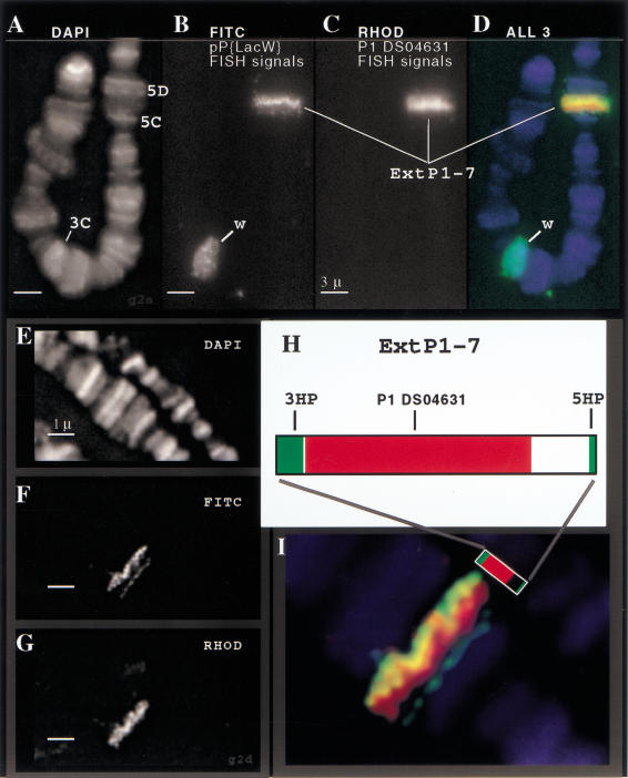
Cytogenetic fine structure FISH analysis of ExtP1-7. Direct-labeled FISH probes were used to visualize the extended P element, ExtP1-7, on the X chromosome. Three-color staining allowed simultaneous detection of chromatin (DAPI images, panels A,E), the boundaries of the extended P elements (pP{LacW} FISH signals—FITC channel, panels B,F), and the interior portion of the extended P (P1-DS04361 FISH signals—rhodamine channel, panels C,G). A close-up view of the pseudocolored overlay image (panel I) reveals the cytogenetic fine structure of the extended P element. The FISH images show good correspondence between the FISH staining patterns (I) and the structure of the extended P element (H; see Fig. 1).
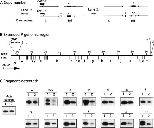
Assessment of structural integrity of the X-linked extended P element ExtP1-7 by quantitative Southern analysis. (A) Genotypes of fly strains used for genomic copy-number comparison, shown with respect to the copy number of the genomic region encompassed by the extended P element (shown as a solid black bar) on the X chromosome (for ExtP1-7) and on the third chromosome. (B) The EcoRI restriction map of the genomic region encompassed by the extended P element. Genomic DNA is shown as a solid line with size kb above and the EcoRI restriction sites below. Specific EcoRI restriction fragments used for the copy-number comparison are labeled with bold letters a–m (Spradling 1981) and w–z. (C) Representative quantitative Southern hybridization results are shown for 16 EcoRI restriction fragments that were identified with 12 independent probes.
Similar articles
-
Systematic gene targeting on the X chromosome of Drosophila melanogaster.Chromosoma. 2004 Dec;113(6):271-5. doi: 10.1007/s00412-004-0313-5. Epub 2004 Oct 12. Chromosoma. 2004. PMID: 15480728
-
Comparison of two active HeT-A retroposons of Drosophila melanogaster.Chromosoma. 1994 Apr;103(2):90-8. doi: 10.1007/BF00352317. Chromosoma. 1994. PMID: 8055715
-
Characterization of MR (P) strains of Drosophila melanogaster: the number of intact P elements and their genetic effect.Genet Res. 1991 Dec;58(3):211-23. doi: 10.1017/s0016672300029967. Genet Res. 1991. PMID: 1666390
-
Gross chromosome rearrangements mediated by transposable elements in Drosophila melanogaster.Bioessays. 1994 Apr;16(4):269-75. doi: 10.1002/bies.950160410. Bioessays. 1994. PMID: 8031304 Review.
-
Insertional mutagenesis of the Drosophila genome with single P elements.Science. 1988 Mar 4;239(4844):1121-8. doi: 10.1126/science.2830671. Science. 1988. PMID: 2830671 Review.
Cited by
-
Technology transfer from worms and flies to vertebrates: transposition-based genome manipulations and their future perspectives.Genome Biol. 2007;8 Suppl 1(Suppl 1):S1. doi: 10.1186/gb-2007-8-s1-s1. Genome Biol. 2007. PMID: 18047686 Free PMC article. Review.
-
Giant Transposons in Eukaryotes: Is Bigger Better?Genome Biol Evol. 2019 Mar 1;11(3):906-918. doi: 10.1093/gbe/evz041. Genome Biol Evol. 2019. PMID: 30796812 Free PMC article. Review.
References
-
- Ashburner M. Drosophila: a laboratory manual. Cold Spring Harbor: Cold Spring Harbor Laboratory; 1989.
-
- Bass HW, Riera-Lizarazu O, Ananiev EV, Bordoli SJ, Rines HW, Phillips RL, Sedat JW, Agard DA, Cande WZ. Evidence for the coincident initiation of homolog pairing and synapsis during the telomere-clustering (bouquet) stage of meiotic prophase. J Cell Sci. 2000;113:1033–1042. - PubMed
-
- Beall EL, Rio DC. Drosophila IRBP/Ku p70 corresponds to the mutagen-sensitive mus309 gene and is involved in P-element excision in vivo. Genes & Dev. 1996;10:921–933. - PubMed
-
- Bier E, Jan LY, Jan YN. rhomboid, a gene required for dorsoventral axis establishment and peripheral nervous system development in Drosophila melanogaster. Genes & Dev. 1990;4:190–203. - PubMed
Publication types
MeSH terms
Substances
Grants and funding
LinkOut - more resources
Full Text Sources
Molecular Biology Databases