. 2000 Nov;74(22):10535-50.
doi: 10.1128/jvi.74.22.10535-10550.2000.

Intracellular localization of vaccinia virus extracellular enveloped virus envelope proteins individually expressed using a Semliki Forest virus replicon

Affiliations

Intracellular localization of vaccinia virus extracellular enveloped virus envelope proteins individually expressed using a Semliki Forest virus replicon

M M Lorenzo et al. J Virol. 2000 Nov.

Abstract

The extracellular enveloped virus (EEV) form of vaccinia virus is bound by an envelope which is acquired by wrapping of intracellular virus particles with cytoplasmic vesicles containing trans-Golgi network markers. Six virus-encoded proteins have been reported as components of the EEV envelope. Of these, four proteins (A33R, A34R, A56R, and B5R) are glycoproteins, one (A36R) is a nonglycosylated transmembrane protein, and one (F13L) is a palmitylated peripheral membrane protein. During infection, these proteins localize to the Golgi complex, where they are incorporated into infectious virus that is then transported and released into the extracellular medium. We have investigated the fates of these proteins after expressing them individually in the absence of vaccinia infection, using a Semliki Forest virus expression system. Significant amounts of proteins A33R and A56R efficiently reached the cell surface, suggesting that they do not contain retention signals for intracellular compartments. In contrast, proteins A34R and F13L were retained intracellularly but showed distributions different from that of the normal infection. Protein A36R was partially retained intracellularly, decorating both the Golgi complex and structures associated with actin fibers. A36R was also transported to the plasma membrane, where it accumulated at the tips of cell projections. Protein B5R was efficiently targeted to the Golgi region. A green fluorescent protein fusion with the last 42 C-terminal amino acids of B5R was sufficient to target the chimeric protein to the Golgi region. However, B5R-deficient vaccinia virus showed a normal localization pattern for other EEV envelope proteins. These results point to the transmembrane or cytosolic domain of B5R protein as one, but not the only, determinant of the retention of EEV proteins in the wrapping compartment.


Figures

FIG. 1
FIG. 1
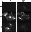
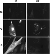
Distribution of vaccinia virus EEV envelope proteins. Immunofluorescence staining was performed on nonpermeabilized cells (NP) and Triton X-100-permeabilized cells (P). Note the bright central staining in permeabilized cells, which corresponds to the Golgi complex area.
FIG. 2
FIG. 2
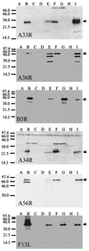
Western blots of proteins expressed using SFV replicons. For each protein, Western blotting was carried out on cell extracts from mock-infected cells (lanes A), cells infected with vaccinia virus WR (lanes B and F) or vaccinia virus deficient in the corresponding protein (lanes C and G), control SFV expressing GFP (lanes D and H) or SFV expressing the corresponding protein (lanes E and I). Lanes A to E correspond to extracts prepared at 7 h postinfection, and lanes F to I correspond to extracts prepared at 24 h postinfection. Protein molecular weight markers are shown at the left of each panel. The arrow at the right of each panel indicates the position of the full-size protein.
FIG. 3
FIG. 3
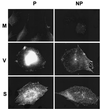
Localization of A33R. Immunofluorescence staining was carried out on BHK-21 cells fixed at 7 h postinfection. Both nonpermeabilized (NP) and Triton X-100-permeabilized (P) cells are shown. The cells were either mock infected (M), infected with vaccinia virus (V), or infected with SFV-A33R particles (S). Note the fine punctate surface staining in cells infected with SFV-A33R.
FIG. 4
FIG. 4
Localization of A33R expressed from SFV-A33R by immunogold staining. Note the high concentration of label on the plasma membrane (arrowheads). Details of membrane labeling of a microvilluslike projection are shown in the inset. Bars, 100 nm.
FIG. 5
FIG. 5
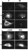
Localization of A34R. (A) Immunofluorescence staining was carried out on BHK-21 cells fixed at 7 h postinfection. Both nonpermeabilized (NP) and Triton X-100-permeabilized (P) cells are shown. The cells were either mock infected (M), infected with vaccinia virus (V), or infected with SFV-A34R particles (S). (B) Costaining with rhodamine-labeled WGA. Cells infected with vaccinia virus (V) or with SFV-A34R (S) were fixed and permeabilized and subjected to WGA and anti-A34R labeling. The same cells are shown on the left and right. Note the localization of A34R in the Golgi region in vaccinia virus-infected cells and the more extended localization in SFV-A34R-infected cells.
FIG. 6
FIG. 6
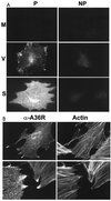
Localization of A36R protein. (A) Immunofluorescence staining was carried out on BHK-21 cells fixed at 7 h postinfection. Both nonpermeabilized (NP) and Triton X-100-permeabilized (P) cells are shown. The cells were either mock infected (M), infected with vaccinia virus (V), or infected with SFV-A36R particles (S). (B) Costaining with rhodamine-labeled phalloidin to reveal actin fibers. Cells infected with SFV-A36R were fixed and permeabilized and subjected to phalloidin and anti-A36R labeling. The same cells are shown on the left and right. Note the localization of A36R in stress fibers and the strong labeling at the tips of cell projections.
FIG. 7
FIG. 7
Immunoelectron microscopy of cells expressing A36R protein. (A) Uniform high labeling of the plasma membrane. There is also some label on the membrane of a putative endosome (E), whereas the mitochondrion (M) is unlabeled. (B) Details of plasma membrane labeling (small arrowheads), including a prominent surface projection (large arrowhead). (C) Heavy labeling for A36R in membrane structures that are in continuity (arrowhead) with the nuclear envelope (N, nucleus). Note the label in vesicles close to the nucleus and in the nuclear envelope (arrows). (D) Significant labeling (arrows) on one side of the Golgi stacks (G). The arrowheads indicate putative clathrin-coated vesicles, whose presence is an indication that the labeled aspect of the Golgi is the TGN. Bars, 100 nm.
FIG. 7
FIG. 7
Immunoelectron microscopy of cells expressing A36R protein. (A) Uniform high labeling of the plasma membrane. There is also some label on the membrane of a putative endosome (E), whereas the mitochondrion (M) is unlabeled. (B) Details of plasma membrane labeling (small arrowheads), including a prominent surface projection (large arrowhead). (C) Heavy labeling for A36R in membrane structures that are in continuity (arrowhead) with the nuclear envelope (N, nucleus). Note the label in vesicles close to the nucleus and in the nuclear envelope (arrows). (D) Significant labeling (arrows) on one side of the Golgi stacks (G). The arrowheads indicate putative clathrin-coated vesicles, whose presence is an indication that the labeled aspect of the Golgi is the TGN. Bars, 100 nm.
FIG. 8
FIG. 8
Localization of A56R. Immunofluorescence staining was carried out on BHK-21 cells fixed at 7 h postinfection. Both nonpermeabilized (NP) and Triton X-100-permeabilized (P) cells are shown. The cells were either mock infected (M), infected with vaccinia virus (V), or infected with SFV-A56R particles (S). Note the strong labeling of the plasma membrane in both vaccinia virus- and SFV-A56R-infected cells.
FIG. 9
FIG. 9
Localization of B5R protein. (A) Immunofluorescence staining was carried out on BHK-21 cells fixed at 7 h postinfection. Both nonpermeabilized (NP) and Triton X-100-permeabilized (P) cells are shown. The cells were either mock infected (M), infected with vaccinia virus (V), or infected with SFV-B5R particles (S). (B) Costaining with rhodamine-labeled WGA. Cells infected with vaccinia virus (V) or SFV-B5R (S) were fixed and permeabilized and subjected to WGA and anti-B5R labeling. The same cells are shown on the right and left. Note that localization of B5R is coincident with the Golgi region in both vaccinia virus-infected cells and SFV-B5R-infected cells.
FIG. 9
FIG. 9
Localization of B5R protein. (A) Immunofluorescence staining was carried out on BHK-21 cells fixed at 7 h postinfection. Both nonpermeabilized (NP) and Triton X-100-permeabilized (P) cells are shown. The cells were either mock infected (M), infected with vaccinia virus (V), or infected with SFV-B5R particles (S). (B) Costaining with rhodamine-labeled WGA. Cells infected with vaccinia virus (V) or SFV-B5R (S) were fixed and permeabilized and subjected to WGA and anti-B5R labeling. The same cells are shown on the right and left. Note that localization of B5R is coincident with the Golgi region in both vaccinia virus-infected cells and SFV-B5R-infected cells.
FIG. 10
FIG. 10
Immunoelectron microscopy of cells expressing B5R protein. (A) Overview of labeling, with prominently labeled intracellular vesicles (arrows). There is little labeling on the plasma membrane (P), and one gold particle is seen over the nuclear envelope (N, nucleus). (B and C) Details of the vesicle labeling. Bars, 100 nm.
FIG. 11
FIG. 11
Localization of F13L protein. Immunofluorescence staining was carried out on BHK-21 cells fixed at 7 h postinfection. Both nonpermeabilized (NP) and Triton X-100-permeabilized (P) cells are shown. The cells were either mock infected (M), infected with vaccinia virus (V), or infected with SFV-F13L particles (S).
FIG. 12
FIG. 12
Localization of B5R protein chimeras. Cells infected with the indicated vaccinia viruses (WR-GFP or WR-GFPmB5R) or with the corresponding SFV replicons (SFV-GFP or SFV-GFPmB5R) are shown.
FIG. 13
FIG. 13
Localization of EEV envelope proteins in vaccinia virus B5R mutants. Cells infected with the indicated vaccinia viruses were fixed and stained with antibody to F13L protein. Wild-type virus (WR) or mutant viruses lacking most of the B5R gene (W-B5R), most of the extracellular domain (W-B5RΔSCR1-4), or the cytoplasmic tail (W-B5RΔc) were used. Note the juxtanuclear localization of F13L in all cases. Differences in the number of enveloped virions reflect differences caused by B5R mutation.

Similar articles

Cited by

References

    1. Appleyard G, Andrews C. Neutralizing activities of antisera to poxvirus soluble antigens. J Gen Virol. 1974;23:197–200. - PubMed
    1. Appleyard G, Hapel A J, Boulter E A. An antigenic difference between intracellular and extracellular rabbitpox virus. J Gen Virol. 1971;13:9–17. - PubMed
    1. Berglund P, Sjoberg M, Garoff H, Atkins G J, Sheahan B J, Liljestrom P. Semliki Forest virus expression system: production of conditionally infectious recombinant particles. Bio/Technology. 1993;11:916–920. - PubMed
    1. Blasco R, Moss B. Extracellular vaccinia virus formation and cell-to-cell virus transmission are prevented by deletion of the gene encoding the 37,000-dalton outer envelope protein. J Virol. 1991;65:5910–5920. - PMC - PubMed
    1. Blasco R, Moss B. Role of cell-associated enveloped vaccinia virus in cell-to-cell virus spread. J Virol. 1992;66:4170–4179. - PMC - PubMed

Publication types

Substances

LinkOut - more resources