. 2000 Nov;74(22):10589-99.
doi: 10.1128/jvi.74.22.10589-10599.2000.

Development of multigene and regulated lentivirus vectors

Affiliations

Development of multigene and regulated lentivirus vectors

J Reiser et al. J Virol. 2000 Nov.

Abstract

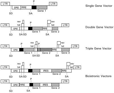
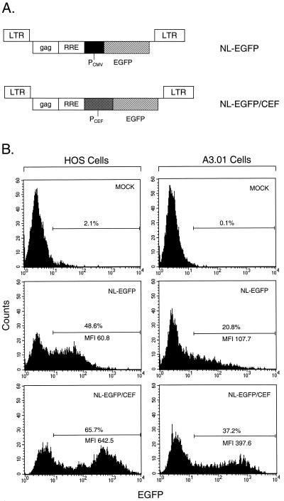
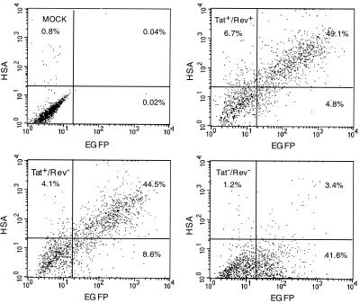
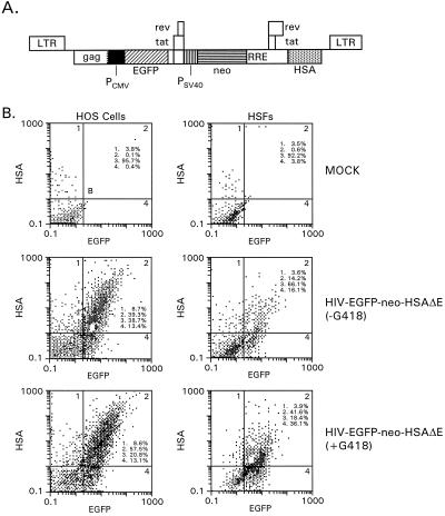
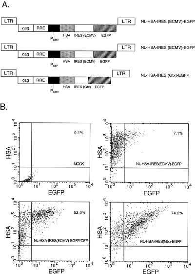
Previously we described safe and efficient three-component human immunodeficiency virus type 1 (HIV-1)-based gene transfer systems for delivery of genes into nondividing cells (H. Mochizuki, J. P. Schwartz, K. Tanaka, R. O. Brady, and J. Reiser, J. Virol. 72:8873-8883, 1998). To apply such vectors in anti-HIV gene therapy strategies and to express multiple proteins in single target cells, we have engineered HIV-1 vectors for the concurrent expression of multiple transgenes. Single-gene vectors, bicistronic vectors, and multigene vectors expressing up to three exogenous genes under the control of two or three different transcriptional units, placed within the viral gag-pol coding region and/or the viral nef and env genes, were designed. The genes encoding the enhanced version of green fluorescent protein (EGFP), mouse heat-stable antigen (HSA), and bacterial neomycin phosphotransferase were used as models whose expression was detected by fluorescence-activated cell sorting, fluorescence microscopy, and G418 selection. Coexpression of these reporter genes in contact-inhibited primary human skin fibroblasts (HSFs) persisted for at least 6 weeks in culture. Coexpression of the HSA and EGFP reporter genes was also achieved following cotransduction of target cells using two separate lentivirus vectors encoding HSA and EGFP, respectively. For the regulated expression of transgenes, tetracycline (Tet)-regulatable lentivirus vectors encoding the reverse Tet transactivator (rtTA) and EGFP controlled by a Tet-responsive element (TRE) were constructed. A binary HIV-1-based vector system consisting of a lentivirus encoding rtTA and a second lentivirus harboring a TRE driving the EGFP reporter gene was also designed. Doxycycline-modulated expression of the EGFP transgene was confirmed in transduced primary HSFs. These versatile vectors can potentially be used in a wide range of gene therapy applications.


Figures

FIG. 1
FIG. 1
HIV-1-based gene transfer vectors. Boxes interrupted by jagged lines contain partial deletions. Abbreviations: P, heterologous transcription promoter; SD, splice donor site; SA, splice acceptor site.
FIG. 2
FIG. 2
Influence of internal promoters on expression of EGFP transgene in HOS cells and in A3.01 T cells. (A) NL-EGFP and NL-EGFP/CEF single-gene vector constructs harboring CMV IE and CEF promoters, respectively. PCMV, human CMV IE promoter; PCEF, hybrid promoter consisting of the enhancer region of the CMV IE promoter fused to translation elongation factor 1α promoter elements. (B) FACS analysis of transduced HOS cells and A3.01 cells. HOS cells were transduced with NL-EGFP and NL-EGFP/CEF vector stocks at MOIs of 0.20 and 0.22, respectively, in DMEM–10% FBS containing Polybrene at 8 μg/ml. A3.01 cells were transduced with the NL-EGFP and NL-EGFP/CEF vector stocks at MOIs of 8.4 and 9.4, respectively, in RPMI 1640 medium–10% FBS containing Polybrene at 8 μg/ml. Cells were incubated with unconcentrated viral supernatants for 18.5 h at 37°C. Forty-eight hours later, the cells were subjected to single-color FACS analysis.
FIG. 3
FIG. 3
Coexpression of EGFP and HSA transgenes following cotransduction of HOS cells with NL-EGFP and NL-HSA vectors. NL-EGFP virus (upper right panel) and NL-HSA virus (lower left panel) were used at MOIs of 0.55 and 0.52, respectively. Cotransduction was carried out using a mixture of NL-EGFP and NL-HSA vector stocks (lower right panel) at the MOIs indicated above. The cells were incubated with virus supernatant for 18 h at 37°C. Forty-eight hours later, cells were detached using trypsin-EDTA and then incubated with a PE-labeled anti-HSA monoclonal antibody (5-μg/ml final concentration) in a total volume of 0.3 ml for 30 min on ice, washed twice with Hanks–2% FBS and then subjected to double-color FACS analysis.
FIG. 4
FIG. 4
Analysis of HOS cells and contact-inhibited primary HSFs following transduction with Tat-dependent double-gene vectors. (A) Vector construct. An EGFP expression cassette consisting of EGFP sequences and the CMV IE promoter was inserted within the viral env coding region. HSA sequences were inserted at the 5′ end of nef. (B) FACS analysis of transduced cells. HOS cells in six-well plates were incubated with HIV-EGFP-HSAΔE and HIV-EGFP-HSAΔE tat(−) vector stocks for 5.5 h at MOIs of 0.65 and 0.67, respectively. Three days after transduction, the cells were detached and then incubated with a PE-labeled anti-HSA monoclonal antibody (5-μg/ml final concentration) in a total volume of 0.3 ml for 30 min on ice. The cells were washed twice and then subjected to double-color FACS analysis. (C) Analysis of transduced HSFs by fluorescence microscopy. Contact-inhibited HSFs on coverslips were incubated with HIV-EGFP-HSAΔE and HIV-EGFP-HSAΔE tat(−) vector stocks for 5.5 h at MOIs of 5.0 and 2.2, respectively. Twenty-eight days later, the cells were stained with a PE-labeled anti-HSA monoclonal antibody (0.4-μg/ml final concentration) and processed for fluorescence microscopy. Top: HSFs transduced with a vector containing functional Tat. Bottom: HSFs transduced with a vector lacking functional Tat. HSA-positive cells are red, and EGFP-positive cells are green.
FIG. 4
FIG. 4
Analysis of HOS cells and contact-inhibited primary HSFs following transduction with Tat-dependent double-gene vectors. (A) Vector construct. An EGFP expression cassette consisting of EGFP sequences and the CMV IE promoter was inserted within the viral env coding region. HSA sequences were inserted at the 5′ end of nef. (B) FACS analysis of transduced cells. HOS cells in six-well plates were incubated with HIV-EGFP-HSAΔE and HIV-EGFP-HSAΔE tat(−) vector stocks for 5.5 h at MOIs of 0.65 and 0.67, respectively. Three days after transduction, the cells were detached and then incubated with a PE-labeled anti-HSA monoclonal antibody (5-μg/ml final concentration) in a total volume of 0.3 ml for 30 min on ice. The cells were washed twice and then subjected to double-color FACS analysis. (C) Analysis of transduced HSFs by fluorescence microscopy. Contact-inhibited HSFs on coverslips were incubated with HIV-EGFP-HSAΔE and HIV-EGFP-HSAΔE tat(−) vector stocks for 5.5 h at MOIs of 5.0 and 2.2, respectively. Twenty-eight days later, the cells were stained with a PE-labeled anti-HSA monoclonal antibody (0.4-μg/ml final concentration) and processed for fluorescence microscopy. Top: HSFs transduced with a vector containing functional Tat. Bottom: HSFs transduced with a vector lacking functional Tat. HSA-positive cells are red, and EGFP-positive cells are green.
FIG. 5
FIG. 5
FACS analysis of HOS cells following transduction with a double-gene vector lacking functional Rev. Cells were incubated with HIV-EGFP-HSAΔE, HIV-EGFP-HSAΔE rev(−), and HIV-EGFP-HSAΔE tat(−)/rev(−) vector stocks for 14 h at MOIs of 1.1, 1.3, and 1.0, respectively. Forty-six hours later, the cells were detached and then incubated with a PE-labeled anti-HSA monoclonal antibody (2.5-μg/ml final concentration) in a total volume of 0.2 ml for 30 min on ice. The cells were washed twice and then subjected to double-color FACS analysis. Upper left, mock-transduced cells (using a HIV-neoΔE vector stock); upper right, cells transduced with the HIV-EGFP-HSAΔE vector; lower left, cells transduced with the HIV-EGFP-HSAΔE rev(−) vector stock; lower right, cells transduced with the HIV-EGFP-HSAΔE tat(−)/rev(−) vector stock.
FIG. 6
FIG. 6
Analysis of HOS cells and contact-inhibited primary HSFs following transduction with a triple-gene vector. (A) Vector construct. An EGFP expression cassette consisting of EGFP sequences and the CMV IE promoter was inserted within the viral gag-pol coding region. A second expression cassette consisting of neo sequences driven by the SV40 early promoter was placed within the env coding region. HSA sequences were inserted at the 5′ end of nef. (B) FACS analysis of transduced HOS cells and HSFs. Top, mock-infected cells; middle, cells grown in the absence of G418; bottom, cells grown in the presence of G418. HOS cells were incubated for 7.5 h with unconcentrated virus at an MOI of 1.3. Contact-inhibited primary HSFs were kept in culture for 27 days prior to transduction. Cells were incubated with unconcentrated virus for 7.5 h using an MOI of 1.1. Three days later, the cells were removed from the wells with trypsin-EDTA and diluted into medium with or without G418 (0.3- to 0.4-mg/ml final concentration). HOS cells were diluted at a ratio of 1:2, and HSFs were diluted at a ratio of 1:4. HOS cells were split one more time at a ratio of 1:10 10 days later and processed for FACS analysis after 6 more days. HSFs were processed for FACS analysis 39 days after the first transfer. The cells were stained and processed for FACS analysis as described in the legend to Fig. 4.
FIG. 7
FIG. 7
FACS analysis of HOS and HT1080 cells following transduction with a bicistronic HIV-1 vector. (A) Vector construct containing EGFP and HSA reporter genes linked by the ECMV IRES. (B) FACS analysis of transduced HOS and HT1080 cells. HOS cells were incubated with virus at an MOI of 0.86 (HSA units) for 7 h at 37°C. HT1080 cells were incubated with the virus for 5.5 h at an MOI of 0.61 (HSA units). The cells were processed for FACS analysis 3 days later as described in the legend to Fig. 4.
FIG. 8
FIG. 8
Effects of internal promoter and IRES on efficiency of expression of downstream cistron in a minimal bicistronic HIV-1 vector. (A) Vector constructs containing the CMV IE or CEF promoter and an ECMV or Gtx IRES element. (B) FACS analysis of transduced HOS cells. Upper left, mock-transduced cells; upper right, cells transduced with the NL-HSA-IRES (ECMV)-EGFP vector (MOI, 3.6); lower left, cells transduced with the NL-HSA-IRES (ECMV)-EGFP/CEF vector (MOI, 3.9); lower right, cells transduced with the NL-HSA-IRES (Gtx)-EGFP vector (MOI, 3.7). Cells were incubated for 24 h at 37°C with the various virus stocks at the indicated MOIs (HSA units) and stained and processed for FACS analysis 2 days later as described in the legend to Figure 4.
FIG. 9
FIG. 9
Tetracycline-modulated HIV-1 vectors. (A) Vector constructs. Top, NL-rtTA/TRE-EGFP vector carrying rtTA sequences driven by the CMV IE promoter and EGFP reporter gene sequences driven by a TRE. Bottom, binary vector system consisting of the NL-rtTA vector encoding rtTA and a second vector carrying the EGFP reporter gene driven by the TRE promoter. (B) Fluorescence microscopy of transduced HSFs. HSFs were transduced sequentially with the NL-TRE-EGFP and NL-rtTA vectors. The cells were grown in the absence or presence of DOX (1 μg/ml) for 6 days. (C) Quantitation of EGFP gene expression in HSFs using confocal fluorescence microscopy. Groups: 1, mock-transduced HSFs; 2 and 3, HSFs transduced with the NL-rtTA/TRE-EGFP vector grown in the absence (group 2) or presence (group 3) of DOX; 4 and 5, HSFs transduced with the binary vector system grown in the absence (group 4) or presence (group 5) of DOX. Cells were incubated with the vector stocks for 22 h in the presence or absence of DOX at MOIs of 2.7 (single-vector system) and 4.0 (binary system). The fluorescence of intracellular EGFP was quantitated by confocal laser scanning fluorescence microscopy (Leica TCS4D). The fluorescence intensity of individual cells was measured using Leica quantitation software. For each group, the relative intensity of three or more typical cells was measured and the mean fluorescence per unit area of the cell was calculated.
FIG. 9
FIG. 9
Tetracycline-modulated HIV-1 vectors. (A) Vector constructs. Top, NL-rtTA/TRE-EGFP vector carrying rtTA sequences driven by the CMV IE promoter and EGFP reporter gene sequences driven by a TRE. Bottom, binary vector system consisting of the NL-rtTA vector encoding rtTA and a second vector carrying the EGFP reporter gene driven by the TRE promoter. (B) Fluorescence microscopy of transduced HSFs. HSFs were transduced sequentially with the NL-TRE-EGFP and NL-rtTA vectors. The cells were grown in the absence or presence of DOX (1 μg/ml) for 6 days. (C) Quantitation of EGFP gene expression in HSFs using confocal fluorescence microscopy. Groups: 1, mock-transduced HSFs; 2 and 3, HSFs transduced with the NL-rtTA/TRE-EGFP vector grown in the absence (group 2) or presence (group 3) of DOX; 4 and 5, HSFs transduced with the binary vector system grown in the absence (group 4) or presence (group 5) of DOX. Cells were incubated with the vector stocks for 22 h in the presence or absence of DOX at MOIs of 2.7 (single-vector system) and 4.0 (binary system). The fluorescence of intracellular EGFP was quantitated by confocal laser scanning fluorescence microscopy (Leica TCS4D). The fluorescence intensity of individual cells was measured using Leica quantitation software. For each group, the relative intensity of three or more typical cells was measured and the mean fluorescence per unit area of the cell was calculated.

References

    1. Adachi A, Gendelman H E, Koenig S, Folks T, Willey R, Rabson A, Martin M A. Production of acquired immunodeficiency syndrome-associated retrovirus in human and nonhuman cells transfected with an infectious molecular clone. J Virol. 1986;59:284–291. - PMC - PubMed
    1. Akkina R K, Walton R M, Chen M L, Li Q-X, Planelles V, Chen I S Y. High-efficiency gene transfer into CD34+ cells with a human immunodeficiency virus type 1-based retroviral vector pseudotyped with vesicular stomatitis virus envelope glycoprotein G. J Virol. 1996;70:2581–2585. - PMC - PubMed
    1. An D S, Morizono K, Li Q-X, Mao S H, Lu S, Chen I S Y. An inducible human immunodeficiency virus type 1 (HIV-1) vector which effectively suppresses HIV-1 replication. J Virol. 1999;73:7671–7677. - PMC - PubMed
    1. Blömer U, Kafri T, Randolph-Moore L, Verma I M, Gage F H. Bcl-xL protects adult septal cholinergic neurons from axotomized cell death. Proc Natl Acad Sci USA. 1998;95:2603–2608. - PMC - PubMed
    1. Blömer U, Naldini L, Kafri T, Trono D, Verma I M, Gage F H. Highly efficient and sustained gene transfer in adult neurons with a lentivirus vector. J Virol. 1997;71:6641–6649. - PMC - PubMed

LinkOut - more resources