. 2000 Nov;74(22):10623-30.
doi: 10.1128/jvi.74.22.10623-10630.2000.

Expression of the two major envelope proteins of equine arteritis virus as a heterodimer is necessary for induction of neutralizing antibodies in mice immunized with recombinant Venezuelan equine encephalitis virus replicon particles

Affiliations

Expression of the two major envelope proteins of equine arteritis virus as a heterodimer is necessary for induction of neutralizing antibodies in mice immunized with recombinant Venezuelan equine encephalitis virus replicon particles

U B Balasuriya et al. J Virol. 2000 Nov.

Abstract

RNA replicon particles derived from a vaccine strain of Venezuelan equine encephalitis virus (VEE) were used as a vector for expression of the major envelope proteins (G(L) and M) of equine arteritis virus (EAV), both individually and in heterodimer form (G(L)/M). Open reading frame 5 (ORF5) encodes the G(L) protein, which expresses the known neutralizing determinants of EAV (U. B. R. Balasuriya, J. F. Patton, P. V. Rossitto, P. J. Timoney, W. H. McCollum, and N. J. MacLachlan, Virology 232:114-128, 1997). ORF5 and ORF6 (which encodes the M protein) of EAV were cloned into two different VEE replicon vectors that contained either one or two 26S subgenomic mRNA promoters. These replicon RNAs were packaged into VEE replicon particles by VEE capsid protein and glycoproteins supplied in trans in cells that were coelectroporated with replicon and helper RNAs. The immunogenicity of individual replicon particle preparations (pVR21-G(L), pVR21-M, and pVR100-G(L)/M) in BALB/c mice was determined. All mice developed antibodies against the recombinant proteins with which they were immunized, but only the mice inoculated with replicon particles expressing the G(L)/M heterodimer developed antibodies that neutralize EAV. The data further confirmed that authentic posttranslational modification and conformational maturation of the recombinant G(L) protein occur only in the presence of the M protein and that this interaction is necessary for induction of neutralizing antibodies.


Figures

FIG. 1
FIG. 1
Schematic presentation of EAV-VEE replicon and helper RNAs. (A) VEE full-length infectious cDNA clone and in vitro-transcribed infectious RNA. The shaded box and arrow show the position of the T7 RNA polymerase promoter and direction of in vitro transcription, respectively. (B) EAV-VEE replicon RNAs. The GL, M, and GL/M replicon RNAs are similar to full-length RNA except that the ORF encoding the VEE structural proteins in the full-length RNA was replaced with ORF5, ORF6, or ORF5 and -6 of EAV, respectively. ORF5 and -6 were placed under the control of two different 26S promoters in the GL/M replicon. (C) Bipartite VEE helper RNAs. The bipartite helper system consists of two helper RNAs derived from the V3014Δ520-7505 monopartite helper (37). The dashed lines indicate deleted regions of the VEE genome. One RNA expresses the VEE capsid gene, and the second RNA expresses the envelope glycoprotein genes.
FIG. 2
FIG. 2
(A) SDS-PAGE of [35S]methionine-labeled cell lysate of individual EAV-VRP-infected BHK-21 cells (identified as GL, M, and GL/M). (B) Immunoblotting of recombinant proteins and EAV with various GL and M protein-specific antibodies. Antigens in each lane are indicated above the lanes. Lysate of uninfected BHK-21 cells was used as the negative control antigen (CT). Antibody (IgG) used in each lane is indicated above the antigens. The proteins were either treated with glyco F (+) or mock treated (−), as indicated below the lanes. Proteins were also analyzed by SDS-PAGE under reducing (with 50 mM DTT [+]) and nonreducing (without 50 mM DTT [−]) conditions. The proteins and their Mrs are indicated on either side of each panel.
FIG. 3
FIG. 3
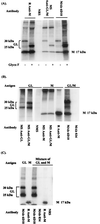
Radiolabeling and immunoprecipitation of EAV envelope proteins expressed from EAV-VRPs in BHK-21 cells by using protein-specific antibodies. The proteins and their Mrs are indicated on either side of each panel. (A) Lysate of pVR100-GL/M-infected cells was immunoprecipitated with antibodies specific for the M protein (rabbit anti-M [R anti-M]) or the GL protein (MAb 6D10) or with sera from mice inoculated with the GL/M heterodimer (mouse anti-GL/M [MS anti-GL/M]) or negative rabbit serum (NRS) and subsequently treated (+) or mock (−) treated with glyco F. (B) Lysates of pVR21-GL-, pVR21-M-, and pVR100-GL/M-infected cells were immunoprecipitated with protein-specific antibodies (R anti-M and MAb 6D10) or sera from mice inoculated with individual EAV-VRPs (e.g., MS anti-GL indicates serum from a mouse immunized with the pVR21-GL VRP) or negative control antibodies (normal mouse serum [NMS] or MAb specific for bluetongue virus [MAb 034]). The antigen (cell lysate) in each lane is identified above the lane, and the antibody used for immunoprecipitation is indicated below the lane. (C) Mixtures of lysates of pVR21-GL- and pVR21-M-infected cells were immunoprecipitated with protein-specific antibodies (R anti-M and MAb 6D10) and negative control antibodies (NRS and MAb 034).
FIG. 4
FIG. 4
Intracellular localization of GL and M proteins in BHK-21 cells infected with EAV-VRPs or EAV (15 h postinoculation). (A) Cells infected with pVR21-GL or pVR21-M are labeled with anti-GL (MAb 6D10)- and rabbit anti-M-purified IgG. The GL protein is stained green with Alexa Flour 488, and the M protein is stained red with Alexa Flour 594 conjugates. (B) Double labeling of pVR100-GL/M-infected cells with anti-GL and anti-M purified IgG. The yellow staining of overlays of both GL and M labels (merged images) indicates that the two proteins colocalize in the Golgi complex when they are present in the same cell and that the GL protein is concentrated in the Golgi complex. (C) The distribution of the GL and M proteins in EAV-infected cells is similar to that in the pVR100-GL/M-infected cells.

References

    1. Balasuriya U B R. Molecular characterization of neutralization determinants and phylogenetic analysis of open reading frame 5 (ORF5) of equine arteritis virus. Ph.D. dissertation. University of California, Davis; 1996.
    1. Balasuriya U B R, MacLachlan N J, de Vries A A F, Rossitto P V, Rottier P J M. Identification of a neutralization site in the major envelope glycoprotein (GL) of equine arteritis virus. Virology. 1995;207:518–527. - PubMed
    1. Balasuriya U B R, Patton J F, Rossitto P V, Timoney P J, McCollum W H, MacLachlan N J. Neutralization determinants of laboratory strains and field isolates of equine arteritis virus: identification of four neutralization sites in the amino-terminal ectodomain of the GL envelope glycoprotein. Virology. 1997;232:114–128. - PubMed
    1. Balasuriya U B R, Rossitto P V, DeMaula C D, MacLachlan N J. A 29K envelope glycoprotein of equine arteritis virus expresses neutralization determinants recognized by murine monoclonal antibodies. J Gen Virol. 1993;74:2525–2529. - PubMed
    1. Balasuriya U B R, Snijder E J, van Dinten L C, Heidner H W, Wilson W D, Hedges J F, Hullinger P J, MacLachlan N J. Equine arteritis virus derived from an infectious cDNA clone is attenuated and genetically stable in infected stallions. Virology. 1999;260:201–208. - PubMed

Publication types

MeSH terms

LinkOut - more resources