. 2000 Nov;74(22):10658-69.
doi: 10.1128/jvi.74.22.10658-10669.2000.

Life cycle of an endogenous retrovirus, ZAM, in Drosophila melanogaster

Affiliations

Life cycle of an endogenous retrovirus, ZAM, in Drosophila melanogaster

P Leblanc et al. J Virol. 2000 Nov.

Abstract

ZAM is an env-containing member of the gypsy family of retrotransposons that represents a possible retrovirus of invertebrates. In this paper, we traced ZAM mobilization to get information about a potential path a retroelement may take to reach the germ line of its host. In situ hybridization on whole-mount tissues and immunocytochemistry analyses with antibodies raised against ZAM Gag and Env proteins have shown that all components necessary to assemble ZAM viral particles, i.e., ZAM full-length RNAs and Gag and Env polypeptides, are coexpressed in a small set of follicle cells surrounding the oocyte. By electron microscopy, we have shown that ZAM viral particles are indeed detected in this somatic lineage of cells, which they leave and enter the closely apposed oocyte. Our data provide evidence that the vesicular traffic and yolk granules in the process of vitellogenesis play an important role in ZAM transfer to the oocyte. Our data support the possibility that vitellogenin transfer to the oocyte may help a retroelement pass to the germ line with no need of its envelope product.


Figures

FIG. 1
FIG. 1
Genetic organization of the ZAM retroelement. The retrovirus-like gag, pol, and env ORFs are flanked by 5′ and 3′ LTRs. The pol and env riboprobes used for in situ hybridization experiments are shown, as are ZAM polypeptides used for preparing polyclonal antibodies directed against Env and Gag proteins (pEnv and pGag, respectively). 1 and 2, oligonucleotides used for the PCR amplification of the gag ORF. The BglII and DraI restriction sites in the env ORF are those used for subcloning the envelope-encoding region into the pRSETC expression vector.
FIG. 2
FIG. 2
In situ hybridization to whole-mount embryos and ovaries to visualize the distribution of ZAM transcripts. (A) Schematic representation of an adult ovariole. The ovariole is composed here of the germarium (early stages of oogenesis) and later of two follicles in stages 9 and 10. The germ line cell nuclei (nurse cells and oocyte nuclei) are in grey, and the somatic cell nuclei (follicle cell nuclei) are in black. (B to F) In situ hybridizations of ZAM RNAs with the antisense env riboprobe (Fig. 1); (G) similar experiment with a sense env riboprobe. (B) Late embryos (up to 10 h at 20°C) of the RevI strain. Arrowheads, hybridization signals located in gonads. (C) Ovariole of RevI female. Arrowheads, hybridization signals located in the somatic follicular cells at the posterior part of each follicle and in the germarium. (D) Higher magnification of RevI follicles (stage 10). Strong hybridization signals are observed (arrowheads) in the posterior follicular cells. (E) Late embryo of the wIR6 strain. After a long time of revelation, a leaky signal is observed in the gonads (arrowheads). (F) Ovariole of the wIR6 strain. No signal is detected in the follicular cells and the germarium. (G) Negative control of RevI follicle hybridized with the sense env riboprobe. In all panels, the anterior part of an embryo or ovariole is at the left margin.
FIG. 3
FIG. 3
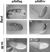
Expression of Gag and Env proteins during Drosophila oogenesis. Shown is the immunolocalization of Gag and Env proteins using polyclonal pAbGag and pAbEnv antibodies, respectively, in ovaries of RevI and wIR6 females. (A to C) Gag of ZAM revealed with the pAbGag antibody in a RevI ovariole. (A) Strong immunostaining is detected in each follicle in a few somatic follicle cells which surround the oocyte and in all follicles. (B) Higher magnification of the posterior part of an early stage 10 follicle. Staining is indicated by arrowheads. (C) Stage 10 follicle from a wIR6 female. No Gag proteins are detected by the pAbGag antibody. (D to F) ZAM Env revealed with the pAbEnv antibody in a RevI ovariole. (D) High level of staining can be observed in a few somatic follicle cells at the posterior part of early stage 10 follicles (arrow). (E) High magnification of the posterior region of the follicle. (F) Stage 10 follicle from a wIR6 female. No Env proteins are detected by the pAbEnv antibody.
FIG. 4
FIG. 4
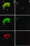
ZAM gag and env genes are coexpressed in the follicle cells of RevI follicles. Double staining of RevI follicles with Gag antibody (red) and Env antibody (green) in early stage 10 of oogenesis (upper panels) and in later stage 10 (lower panels). Bars: upper panels, 10 μm; lower panels, 30 μm. oo, oocyte; fc, follicle cells.
FIG. 5
FIG. 5
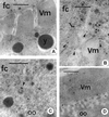
Ultrastructural identification of ZAM viral particles in ovarian follicles of the RevI strain from D. melanogaster. (A) A posterior follicle cell (fc) facing the oocyte (oo) from a stage 9 ovarian follicle is shown. Vm, vitelline membrane. Bar, 0.5 μm. (B) Enlargement of panel A to show the apical end of a follicle cell, where numerous roundish viral particles (arrows) (average diameter, 45 nm) can be clearly seen. Bar, 0.3 μm. (C) The apical end of a posterior follicle cell from a stage 10 ovarian follicle of the wIR6 strain showing several vitelline membrane precursors (pVm) and the vitelline membrane itself, but not viral particles. Bar, 0.9 μm. (Inset) Enlargement of the vitelline membrane precursors.
FIG. 6
FIG. 6
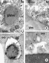
Cytochemical detection of viral particles on vitelline membrane precursors and in yolk granules of the RevI strain. (A) A vitelline membrane precursor (pVm) along the apical end of a posterior follicle cell (fc) from a stage 9 ovarian follicle. Note the presence of numerous viral particles (arrowheads) around the granule periphery. Bar, 0.25 μm. Vm, vitelline membrane. (B) The apical end of a posterior follicle cell from a stage 11 ovarian follicle. Note the presence of numerous viral particles (arrowheads) along the margin of the vitelline membrane. Bar, 0.2 μm. (C) A forming yolk granule (y) from the cortical ooplasm of a stage 9 ovarian follicle of RevI following a 1-h exposure to HRP. Note that viral particles (arrowheads) are present along the superficial layer underneath the limiting membrane. A peroxidase-labeled endocytic vesicle is also visible along the membrane (arrow). oo, oocyte. Bar, 0.5 μm. (D) A forming yolk granule from a stage 9 RevI ovarian follicle fixed for 4 h with zinc osmium iodide (OZI). Note the presence of several viral particles (arrowheads) along the superficial layer among several electron-dense spots of OZI precipitates. Bar, 0.1 μm.
FIG. 7
FIG. 7
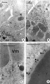
Immunocytochemical detection of Gag viral antigens. (A) The follicle cell-oocyte border from a stage 9 RevI ovarian follicle tested with anti-Gag antibody. fc, posterior follicle cell; N, follicle cell nucleus; oo, oocyte; Vm, vitelline membrane. Bar, 4 μm. (B) Enlargement of panel A to show numerous 20-nm gold grains of the secondary antibody along the apical end of the follicle cell. Bar, 1 μm. (C) Portion of the cortical ooplasm from a stage 10 RevI ovarian follicle showing gold grains (arrowheads) due to anti-Gag antibody along the oolemma. Bar, 0.5 μm. (D) A forming yolk granule (y) from a stage 9 RevI ovarian follicle. Note the presence of gold grains due to anti-Gag antibody (arrowheads) over the superficial layer among viral particles (arrows). Bar, 0.4 μm.
FIG. 8
FIG. 8
Immunocytochemical detection of Env viral antigens. (A) The follicle cell (fc)-oocyte border from a stage 10 RevI ovarian follicle exposed to anti-Env antibody. Gold grains are dispersed over the vitelline membrane (Vm). y, yolk granule. Bar, 0.5 μm. (B) The apical end of a posterior follicle cell from a stage 9 RevI ovarian follicle showing several gold grains (arrowheads) along the plasma membrane. Bar, 0.4 μm. (C) The posterior-most cortical ooplasm from a stage 9 RevI ovarian follicle tested with anti-Env antibody. Arrowhead, gold-labeled coated vesicle. oo, oocyte. Bar, 0.4 μm. (D) Portion of a stage 11 RevI ovarian follicle showing the vitelline membrane and the underneath oolemma. Vitellogenic uptake has ceased by this developmental stage in D. melanogaster, and yet gold grains due to the anti-Env antibody are still seen bound along the microvilli of the oolemma. Bar, 0.6 μm.

Similar articles

Cited by

References

    1. Chalvet F, Teysset L, Terzian C, Prud'Homme N, Santamaria P, Bucheton A, Pélisson A. Proviral amplification of the Gypsy endogenous retrovirus of Drosophila melanogaster involves env-independent invasion of the female germline. EMBO J. 1999;18:2659–2669. - PMC - PubMed
    1. Coffin J M, Hughes S H, Varmus H E. Retroviruses. Cold Spring Harbor, N.Y: Cold Spring Harbor Laboratory Press; 1997. - PubMed
    1. Desset S, Conte C, Dimitri P, Calco V, Dastugue B, Vaury C. Mobilization of two retroelements, ZAM and Idefix, in a novel unstable line of Drosophila melanogaster. Mol Biol Evol. 1999;16:54–66. - PubMed
    1. Dong J, Roth M G, Hunter E. A chimeric avian retrovirus containing the influenza virus hemagglutinin gene has an expanded host range. J Virol. 1992;66:7374–7382. - PMC - PubMed
    1. Friesen P D, Nissen M S. Gene organization and transcription of TED, a lepidopteran retrotransposon integrated within the baculovirus genome. Mol Cell Biol. 1990;10:3067–3077. - PMC - PubMed

Publication types

MeSH terms

LinkOut - more resources