. 2000 Nov;74(22):10778-84.
doi: 10.1128/jvi.74.22.10778-10784.2000.

Lentivirus vector gene expression during ES cell-derived hematopoietic development in vitro

Affiliations

Lentivirus vector gene expression during ES cell-derived hematopoietic development in vitro

I Hamaguchi et al. J Virol. 2000 Nov.

Abstract

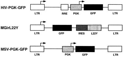
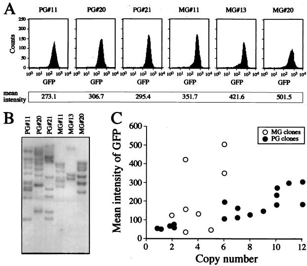
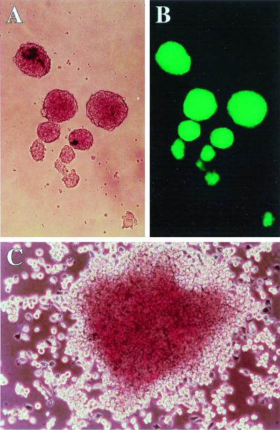
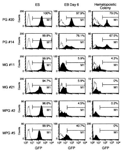
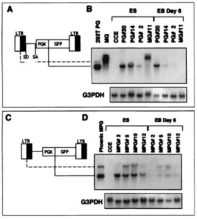
The murine embryonal stem (ES) cell virus (MESV) can express transgenes from the long terminal repeat (LTR) promoter/enhancer in undifferentiated ES cells, but expression is turned off upon differentiation to embryoid bodies (EBs) and hematopoietic cells in vitro. We examined whether a human immunodeficiency virus type 1-based lentivirus vector pseudotyped with the vesicular stomatitis virus G protein (VSV-G) could transduce ES cells efficiently and express the green fluorescent protein (GFP) transgene from an internal phosphoglycerate kinase (PGK) promoter throughout development to hematopoietic cells in vitro. An oncoretrovirus vector containing the MESV LTR and the GFP gene was used for comparison. Fluorescence-activated cell sorting analysis of transduced CCE ES cells showed 99.8 and 86.7% GPF-expressing ES cells in the VSV-G-pseudotyped lentivirus (multiplicity of infection [MOI] = 59)- and oncoretrovirus (MOI = 590)-transduced cells, respectively. Therefore, VSV-G pseudotyping of lentiviral and oncoretrovirus vectors leads to efficient transduction of ES cells. Lentivirus vector integration was verified in the ES cell colonies by Southern blot analysis. When the transduced ES cells were differentiated in vitro, expression from the oncoretrovirus LTR was severely reduced or extinct in day 6 EBs and ES cell-derived hematopoietic colonies. In contrast, many lentivirus-transduced colonies, expressing the GFP gene in the undifferentiated state, continued to express the transgene throughout in vitro development to EBs at day 6, and many continued to express in cells derived from hematopoietic colonies. This experimental system can be used to analyze lentivirus vector design for optimal expression in hematopoietic cells and for gain-of-function experiments during ES cell development in vitro.


Figures

FIG. 1
FIG. 1
Vectors. The lentivirus vector (HIV-PGK-GFP) uses the internal PGK promoter to drive expression of GFP gene. The Rev-responsive element (RRE) is indicated. The oncoretrovirus vectors (MGirL22Y [28]) and MSV-PGK-GFP) contain the same vector backbone using the MESV LTR, the same primer binding site, and the same length of the gag region (the figure is not drawn to scale) (14, 28). MGirL22Y contains the GFP gene followed by an internal ribosomal entry site (IRES) from the encephalomyocarditis virus linked to a mutant dihydrofolate reductase gene (L22Y). MSV-PGK-GFP contains the MESV LTR and the internal PGK promoter followed by the GFP gene.
FIG. 2
FIG. 2
Expression level of GFP and number of integrated proviral copies in transduced ES clones. (A) Three lentivirus (HIV-PGK-GFP vector)- and three oncoretrovirus (MGirL22Y vector)-transduced ES clones (PG and MS clones, respectively) which showed high GFP expression levels. (B) Southern blot analysis of the clones. Genomic DNAs of the PG and MG clones were digested with BamHI and EcoRI, respectively, and hybridized with GFP cDNA. (C) Relationship between the integrated copy number and expression level of GFP.
FIG. 3
FIG. 3
ES-derived differentiated cells. (A and B) Day 6 EBs derived from the lentivirus-transduced clone PG20 (magnification, ×100). Panel B is visualized by a fluorescence microscope. (C) Day 10 hematopoietic colony derived from ES cells (×150).
FIG. 4
FIG. 4
GFP expression in lentivirus- and oncoretrovirus-transduced clones during hematopoietic differentiation. FACS analysis shows the number of GFP-positive cells in undifferentiated ES cells, in day 6 EBs, and in hematopoietic colonies from methylcellulose cultures. Cells from day 6 EBs and hematopoietic colonies were analyzed by gating away dead cells stained with propidium iodide. PG, MG, and MPG indicate clones transduced with HIV-PGK-GFP, MGirL22Y, and MSV-PGK-GFP, respectively.
FIG. 5
FIG. 5
Northern blot analysis of GFP expression in ES clones transduced with lentivirus and oncoretrovirus vectors. Schematic drawings of the lentivirus vector, HIV-PGK-GFP (A), and the retrovirus vector, MSV-PGK-GFP (C), depict the species of RNA generated by the internal promoter (solid line, shorter transcript) and the viral LTR (broken line, longer transcript). The splice donor and acceptor sites (SD and SA) are indicated. (B and D) RNA was extracted from virus-transduced ES cell clones in undifferentiated (ES) and differentiated (EB Day 6) cells. Twenty micrograms of RNA was transferred onto a nylon membrane and hybridized with a radioactively labeled DNA fragment specific for GFP and glyceraldehyde 3-phosphate dehydrogenase (G3PDH). RNA from 293T cells producing HIV-PGK-GFP, the MGirL22Y producer cells, and ecotropic Phoenix cells transfected with the MSV-PGK-GFP vector are shown for comparison. The MG lane in panel B shows two bands of 3.2 kb (unspliced) and 2.5 kb (spliced). In the lentivirus vector clones, a 1.4-kb mRNA species is generated by the PGK promoter. PG, MG, and MPG indicate clones transduced with HIV-PGK-GFP, MGirL22Y, and MSV-PGK-GFP, respectively. CCE, untransduced ES cells.

References

    1. Akkina R K, Walton R M, Chen M L, Li Q X, Planelles V, Chen I S. High-efficiency gene transfer into CD34+ cells with a human immunodeficiency virus type 1-based retroviral vector pseudotyped with vesicular somatitis virus envelope glycoprotein G. J Virol. 1996;70:2581–2585. - PMC - PubMed
    1. Barklis E, Mulligan R C, Jaenisch R. Chromosomal position or virus mutation permits retrovirus expression in embryonal carcinoma cells. Cell. 1986;47:391–399. - PubMed
    1. Bartz S R, Vodicka M A. Production of high-titer human immunodeficiency virus type 1 pseudotyped with vesicular somatitis virus glycoprotein. Methods. 1997;12:337–342. - PubMed
    1. Becker K G, Jedlicka P, Templeton N S, Loitta L, Ozato K. Characterization of hUCRBP(YY1,NF-E1,δ): a transcription factor that binds to the regulatory regions of many viral and cellular genes. Gene. 1994;150:259–266. - PubMed
    1. Blömer U, Naldini L, Kafri T, Trono D, Verma I M, Gage F H. Highly efficient and sustained gene transfer in adult neurons with a lentivirus vector. J Virol. 1997;71:6641–6649. - PMC - PubMed

Publication types

MeSH terms

Substances