. 2000 Nov;20(22):8458-67.
doi: 10.1128/MCB.20.22.8458-8467.2000.

Multiple C-terminal lysine residues target p53 for ubiquitin-proteasome-mediated degradation

Affiliations

Multiple C-terminal lysine residues target p53 for ubiquitin-proteasome-mediated degradation

M S Rodriguez et al. Mol Cell Biol. 2000 Nov.

Abstract

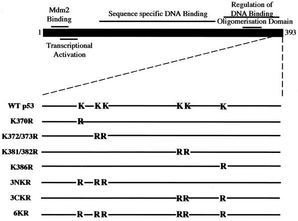
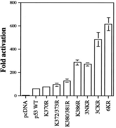
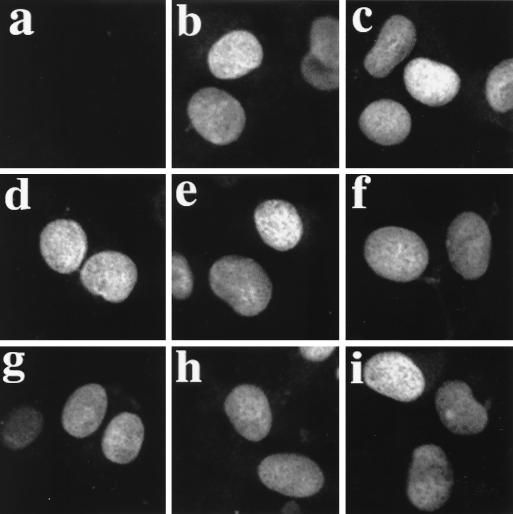
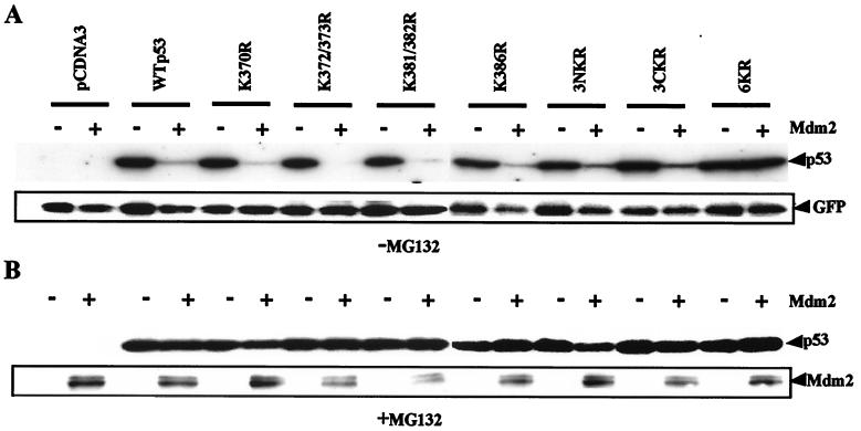
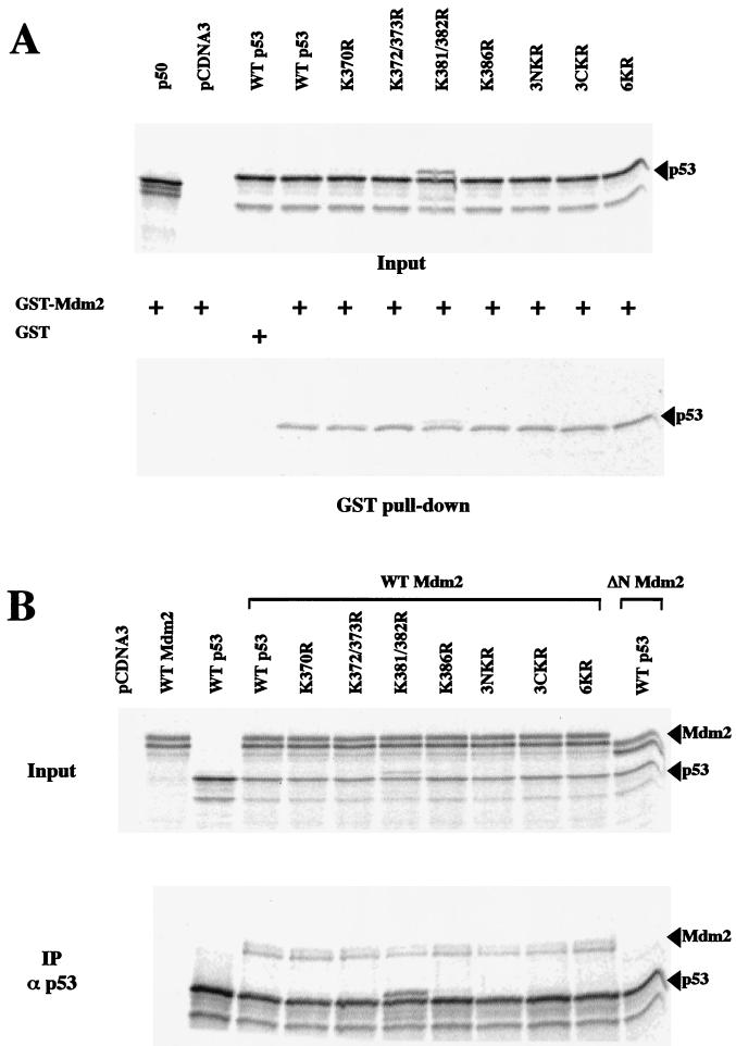
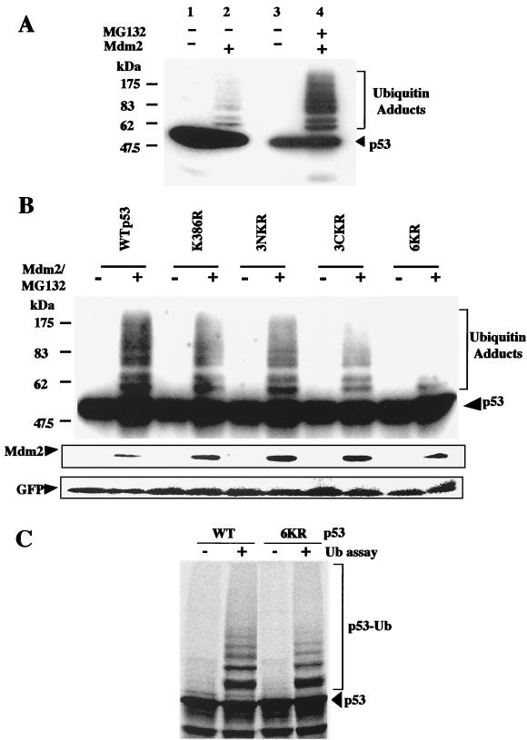
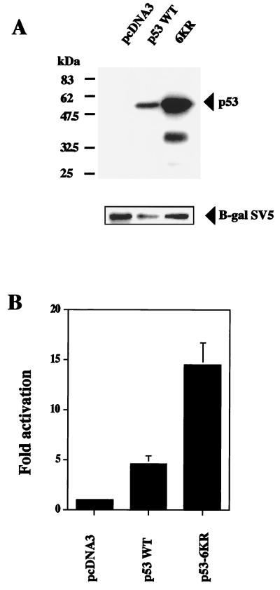
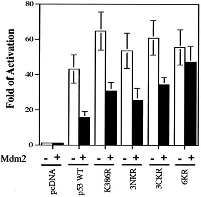
In normal cells, p53 is maintained at a low level by ubiquitin-mediated proteolysis, but after genotoxic insult this process is inhibited and p53 levels rise dramatically. Ubiquitination of p53 requires the ubiquitin-activating enzyme Ubc5 as a ubiquitin conjugation enzyme and Mdm2, which acts as a ubiquitin protein ligase. In addition to the N-terminal region, which is required for interaction with Mdm2, the C-terminal domain of p53 modulates the susceptibility of p53 to Mdm2-mediated degradation. To analyze the role of the C-terminal domain in p53 ubiquitination, we have generated p53 molecules containing single and multiple lysine-to-arginine changes between residues 370 and 386. Although wild-type (WT) and mutant molecules show similar subcellular distributions, the mutants display a higher transcriptional activity than WT p53. Simultaneous mutation of lysine residues 370, 372, 373, 381, 382, and 386 to arginine residues (6KR p53 mutant) generates a p53 molecule with potent transcriptional activity that is resistant to Mdm2-induced degradation and is refractory to Mdm2-mediated ubiquitination. In contrast to WT p53, transcriptional activity directed by the 6KR p53 mutant fails to be negatively regulated by Mdm2. Those differences are also manifest in HeLa cells which express the human papillomavirus E6 protein, suggesting that p53 C-terminal lysine residues are also implicated in E6-AP-mediated ubiquitination. These data suggest that p53 C-terminal lysine residues are the main sites of ubiquitin ligation, which target p53 for proteasome-mediated degradation.


Figures

FIG. 1
FIG. 1
Schematic representation of p53 mutants with C-terminal K-to-R changes.
FIG. 2
FIG. 2
p53 mutants containing C-terminal K-to-R substitutions show higher transcriptional activity than does WT p53. Saos-2 cells were cotransfected by electroporation with expression plasmids for WT p53 or K-to-R mutants and the pG13-Luc reporter plasmid. Each point is the mean of four independent transfections, with error bars representing 1 standard deviation.
FIG. 3
FIG. 3
Subcellular localization of WT p53 and mutants with C-terminal K-to-R changes. H1299 cells were transfected with empty pcDNA3 vector (a) and plasmids expressing WT p53 (b), 6KR (c), 3NKR (d), 3CKR (e), K372/373R (f), K381/382R (g), K370R (h), and K386R (i). Indirect immunofluorescence with antibody against p53 DO.1 is shown.
FIG. 4
FIG. 4
Mdm2-mediated degradation is dependent on lysine residues in the C-terminal region of p53. Saos-2 cells were electroporated with plasmids encoding GFP, WT p53, or K-to-R p53 mutants, together with empty pcDNA3 vector or Mdm2 vector as indicated. (A) Twenty-four hours after transfection, whole-cell extracts were prepared and analyzed by Western blotting with anti-p53, anti-Mdm2, and anti-GFP monoclonal antibodies. (B) Transfected cells were treated with 10 μM MG132 for 4 h prior to harvest. Western blotting against p53 and Mdm2 was performed as described above.
FIG. 5
FIG. 5
K-to-R changes in the p53 C-terminal region do not affect p53-Mdm2 interaction. (A) 35S-labeled NF-κB (p50), WT p53, and K-to-R p53 mutants generated by in vitro transcription and translation were incubated with GST-Mdm2 or GST as indicated. GST pull-down assays were performed at 4°C. Mdm2-associated material was fractionated by SDS-PAGE, and the dried gel was analyzed by phosphorimaging (lower panel). (B) For coimmunoprecipitation, 35S-labeled Mdm2, ΔN Mdm2, WT p53, and K-to-R p53 mutants generated by in vitro transcription and translation were incubated with protein A- and protein G-agarose beads and p53-specific polyclonal antibody CM1, as indicated (IPα p53). p53-associated material was fractionated by SDS-PAGE, and the dried gel was analyzed by phosphorimaging (lower panel).
FIG. 6
FIG. 6
p53 C-terminal lysine residues are targets for Mdm2-mediated ubiquitination. (A) Ubiquitination of p53 in vivo. p53−/− Mdm2−/− cells were cotransfected with WT p53 and empty pcDNA3 vector or an Mdm2 expression plasmid and incubated with proteasome inhibitor MG132, as indicated. Whole-cell extracts were analyzed by Western blotting with DO.1 anti-p53 monoclonal antibody. (B) p53−/− Mdm2−/− cells were cotransfected with a GFP-encoding plasmid and WT p53 or K-to-R mutants, together with empty pcDNA3 vector or an Mdm2 expression plasmid. Cells were treated and analyzed as described above. After being stripped, the membrane was incubated with Mdm2 and GFP antibodies. (C) In vitro ubiquitination (Ub) assays were performed with 35S-labeled WT p53 and the 6KR mutant. Reaction products were fractionated by SDS-PAGE, and the dried gel was analyzed by phosphorimaging. p53-Ub, ubiquitin-conjugated forms of p53.
FIG. 7
FIG. 7
p53 C-terminal lysine residues are targets for E6-AP-mediated degradation. (A) HeLa cells which express HPV E6 protein were cotransfected with WT p53 or the 6KR mutant and SV5-tagged β-galactosidase expression plasmids. Twenty-four hours after transfection, whole-cell extracts were analyzed by Western blotting with a monoclonal antibody to p53 (DO.1). After being stripped, the membrane was blotted with SV5 antibody (β-gal SV5). (B) HeLa cells were cotransfected with the pG13-Luc reporter plasmid and expression plasmids for WT p53 or the 6KR mutant. Twelve hours after transfection, reporter activity was determined. Each point is the mean of four independent transfections, with error bars representing 1 standard deviation.
FIG. 8
FIG. 8
Negative regulation of p53 transcriptional activity by Mdm2 requires ubiquitin-mediated degradation. p53−/− Mdm2−/− cells were cotransfected with the pG13-Luc reporter plasmid and expression plasmids for WT p53 or the 6KR mutant, together with an empty vector or an Mdm2 expression plasmid. Twelve hours after transfection, reporter activity was determined. Each point is the mean of five independent transfections, with error bars representing 1 standard deviation.

Similar articles

Cited by

References

    1. Askjaer P, Jensen T H, Nilsson J, Englmeier L, Kjems J. The specificity of the CRM1-Rev nuclear export signal interaction is mediated by RanGTP. J Biol Chem. 1998;273:33414–33422. - PubMed
    1. Baldi L, Brown K, Franzoso G, Siebenlist U. Critical role for lysine-21 and lysine-22 signal-induced, ubiquitin-mediated proteolysis of IκBα. J Biol Chem. 1996;271:376–379. - PubMed
    1. Barak Y, Juven T, Haffner R, Oren M. mdm2 expression is induced by wild type p53 activity. EMBO J. 1993;12:461–468. - PMC - PubMed
    1. Bottger V, Bottger A, Howard S F, Picksley S M, Chene P, Garcia-Echeverria C, Hochkeppel H K, Lane D P. Identification of novel mdm2 binding peptides by phage display. Oncogene. 1996;13:2141–2147. - PubMed
    1. Chen J, Marechal V, Levine A J. Mapping of the p53 and mdm-2 interaction domains. Mol Cell Biol. 1993;13:4107–4114. - PMC - PubMed

Publication types

MeSH terms

LinkOut - more resources