. 2000 Nov;20(22):8526-35.
doi: 10.1128/MCB.20.22.8526-8535.2000.

Latent membrane protein 2A of Epstein-Barr virus binds WW domain E3 protein-ubiquitin ligases that ubiquitinate B-cell tyrosine kinases

Affiliations

Latent membrane protein 2A of Epstein-Barr virus binds WW domain E3 protein-ubiquitin ligases that ubiquitinate B-cell tyrosine kinases

G Winberg et al. Mol Cell Biol. 2000 Nov.

Abstract

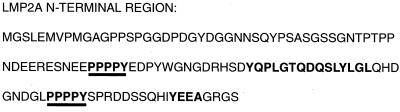
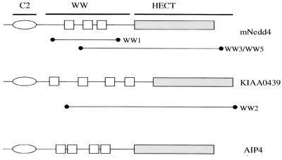
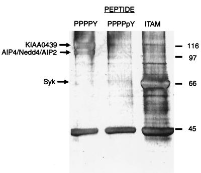
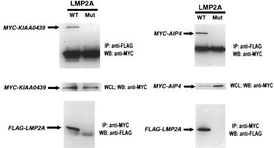
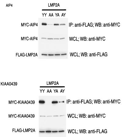
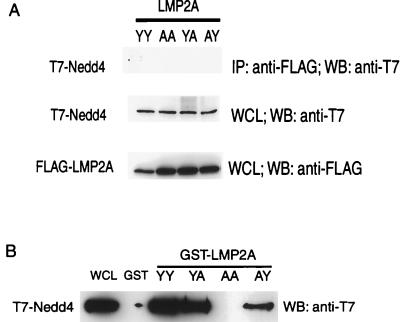
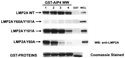
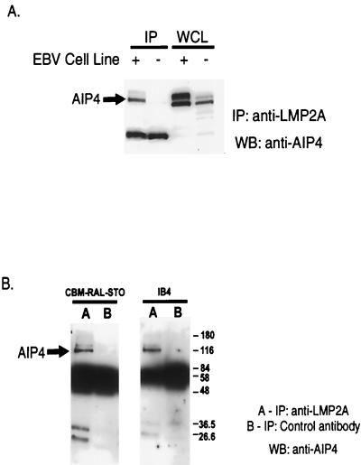
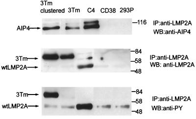
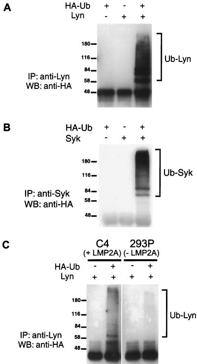
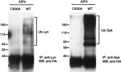
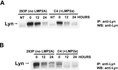
The latent membrane protein (LMP) 2A of Epstein-Barr virus (EBV) is implicated in the maintenance of viral latency and appears to function in part by inhibiting B-cell receptor (BCR) signaling. The N-terminal cytoplasmic region of LMP2A has multiple tyrosine residues that upon phosphorylation bind the SH2 domains of the Syk tyrosine kinase and the Src family kinase Lyn. The LMP2A N-terminal region also has two conserved PPPPY motifs. Here we show that the PPPPY motifs of LMP2A bind multiple WW domains of E3 protein-ubiquitin ligases of the Nedd4 family, including AIP4 and KIAA0439, and demonstrate that AIP4 and KIAA0439 form physiological complexes with LMP2A in EBV-positive B cells. In addition to a C2 domain and four WW domains, these proteins have a C-terminal Hect catalytic domain implicated in the ubiquitination of target proteins. LMP2A enhances Lyn and Syk ubiquitination in vivo in a fashion that depends on the activity of Nedd4 family members and correlates with destabilization of the Lyn tyrosine kinase. These results suggest that LMP2A serves as a molecular scaffold to recruit both B-cell tyrosine kinases and C2/WW/Hect domain E3 protein-ubiquitin ligases. This may promote Lyn and Syk ubiquitination in a fashion that contributes to a block in B-cell signaling. LMP2A may potentiate a normal mechanism by which Nedd4 family E3 enzymes regulate B-cell signaling.


Figures

FIG. 1
FIG. 1
Amino acid sequence of the cytoplasmic N-terminal region of LMP2A. The two PPPPY motifs are underlined in bold type. Also indicated in bold type are the ITAM and YEEA motifs, which, when phosphorylated, become ligands for the B-cell tyrosine kinases Syk and Lyn, respectively.
FIG. 2
FIG. 2
Domain organization of mouse Nedd4, KIAA0439, and AIP4. Also illustrated are the protein fragments of Nedd4 (WW1, WW3, WW5) and KIAA0439 (WW2) that were isolated from a COLT screen of a 10-day mouse embryo expression library probed with the LMP2A-derived synthetic peptide Biotin.Aca.S.N.E.E.P.P.P.P.Y.E.D.P.Y.W.G.N.G.
FIG. 3
FIG. 3
Specific B-cell proteins bind to affinity reagents derived from ligand motifs found in LMP2A. Immobilized LMP2A-derived synthetic peptides based on PPPPY, PPPPpY, and ITAM motifs were treated with clarified lysates from a CBM-RAL-STO B-cell line. Associated proteins were resolved by SDS–9% PAGE, visualized by silver staining, and identified by quadrupole–time-of-flight (mass spectrometry) (Table 1). Numbers are for molecular mass markers (kilodaltons).
FIG. 4
FIG. 4
The E3 ubiquitin-ligase proteins KIAA0439 and AIP4 associate with LMP2A when overexpressed in the B-cell line DG75. A FLAG epitope-tagged LMP2A mammalian expression vector was gene pulsed into DG75 cells with either a MYC epitope-tagged KIAA0439 C962S or AIP4 C830A expression vector. Control experiments using a derivative of LMP2A unable to bind WW domain proteins, due to mutation of both PPPPY motifs to PPPPA, are also shown. Following the gene pulse, the cells were grown for 40 h and then lysed in NP-40 buffer. Lysates were split equally and immunoprecipitated with either anti-MYC or anti-FLAG for 18 h at 5°C. Proteins were resolved by SDS–8% PAGE and subjected to Western blotting for detection of the associated FLAG or MYC epitope-tagged protein. WT, wild type; Mut, mutant; IP, immunoprecipitation; WB, Western blotting; WCL, whole-cell lysak.
FIG. 5
FIG. 5
The LMP2A PPPPY motifs are essential for binding E3 ubiquitin-ligase proteins KIAA0439 and AIP4. Wild-type and PPPPY motif mutants (YY, wild type; AA, Y60A/Y101A; YA, Y101A; AY, Y60A) of a FLAG epitope-tagged LMP2A mammalian expression vector were transfected into Cos-1 cells with either MYC epitope-tagged KIAA0439 or AIP4 expression vectors. Lysates were then subjected to anti-FLAG immunoprecipitations (IP), and associated proteins were identified by SDS–8% PAGE with Western blotting for MYC epitope-tagged protein. WB, Western blotting; WCL, whole-cell lysate.
FIG. 6
FIG. 6
Nedd4 can be precipitated with N-terminal LMP2A GST fusion protein but does not associate with full-length LMP2A when both proteins are overexpressed in Cos-1 cells. (A) Wild type (YY) and PPPPY motif (AA, Y60A/Y101A; YA, Y101A; AY, Y60A) mutants of a FLAG epitope-tagged LMP2A mammalian expression vector were transfected into Cos-1 cells with the T7 epitope-tagged Nedd4 expression vector. Lysates were then subjected to anti-FLAG immunoprecipitations (IP), and associated protein was identified by SDS–8% PAGE with Western blotting (WB) for T7 epitope-tagged protein. (B) Cos-1 cell lysates containing T7 epitope-tagged Nedd4 protein were treated with immobilized GST or GST fusion proteins containing the N-terminal 122 amino acid residues of wild-type LMP2A or PPPPY mutants. Associated Nedd4 was identified by SDS–9% PAGE with Western blotting (WB) for the T7 epitope-tagged protein. WCL, whole-cell lysate.
FIG. 7
FIG. 7
Individual AIP4 WW domains can precipitate LMP2A. Immobilized GST fusion proteins of individual AIP4 WW domains were tested for their ability to precipitate from lysates of wild-type LMP2A (LMP2A WT) or Y60A/Y101A, Y101A, or Y60A LMP2A mutant proteins expressed transiently in DG75 B cells. Western blotting (WB) with anti-LMP2A antibody identified precipitated LMP2A. The numerical designation of each AIP4 WW domain corresponds to its position from the N terminus of the protein. WCL, whole-cell lysate.
FIG. 8
FIG. 8
LMP2A associates with endogenous AIP4 in EBV-positive B-cell lines. (A) LMP2A immunoprecipitations, using rat anti-LMP2A MAb 4E11 from cell lysates of either an EBV-negative (−) cell line, BJAB, or an EBV-positive (+) B-cell line, CBM-RAL-STO, were probed for the presence of AIP4 by Western blotting using anti-AIP4 antibodies. (B) LMP2A immunoprecipitations and control immunoprecipitations using an irrelevant anti-rat MAb IgG1 from cell lysates of two EBV-positive B-cell lines, CBM-RAL-STO and IB4, were probed for AIP4. IP, immunoprecipitation; WB, Western blotting, WCL, whole-cell lysate. Numbers are molecular mass in kilodaltons.
FIG. 9
FIG. 9
The N-terminal cytoplasmic region of LMP2A is sufficient to bind AIP4. Four HEK 293 cell lines that stably express full-length LMP2A (C4), a CD38-LMP2A chimeric molecule (3Tm), a control vector expressing the CD38 portion of the chimeric molecule (CD38), or vector alone (293P) were tested for their ability to form LMP2A-AIP4 complexes with endogenous AIP4. Clustering CD38-LMP2A, through treatment of the cells with anti-CD38 antibody, shows little effect on association with AIP4. However, clustering dramatically increases the level of tyrosine phosphorylation, as determined by immunoblotting with anti-phosphotyrosine (anti-PY) 4G10 antibody. Full-length LMP2A is also tyrosine phosphorylated when expressed in HEK 293 cells. IP, immunoprecipitation; WB, Western blotting; wt, wild type. Numbers are molecular mass in kilodaltons.
FIG. 10
FIG. 10
Lyn and Syk undergo ubiquitination; Lyn ubiquitination is augmented in an LMP2A-expressing cell line. (A) HEK 293 cells were cotransfected with Lyn and HA-ubiquitin expression vectors along with control transfections using each vector separately. Ubiquitinated Lyn species were isolated by immunoprecipitation with anti-Lyn antibody, resolved by SDS–8.3% PAGE, and detected by Western blotting using anti-HA antibody. (B) Ubiquitination of Syk in HEK 293 cells was monitored as described for panel A, with transfection of the Syk expression vector and immunoprecipitation using anti-Syk antibody. (C) Lyn was transfected into either a HEK 293 cell line stably expressing LMP2A (C4) or a control cell line (293P) in the presence or absence of an HA-ubiquitin expression vector. Ubiquitinated Lyn species were isolated by immunoprecipitation with anti-Lyn antibody and detected by Western blotting using anti-HA antibody. Numbers are molecular mass in kilodaltons. IP, immunoprecipitation; WB, Western blotting; Ub, ubiquitin; +, DNA vector added; −, DNA vector not added.
FIG. 11
FIG. 11
An inactive HECT domain form of AIP4 reduces the level of Lyn and Syk ubiquitination in LMP2A-expressing HEK 293 cells. A HEK 293-derived cell line that stably expresses LMP2A (C4) was used for transfection of HA-ubiquitin with either Lyn or Syk expression vectors. Also transfected was wild-type AIP4 (WT) or an inactive form of AIP4 (C830A). The presence of ubiquitinated Lyn (Ub-Lyn) or Syk (Ub-Syk) was detected by immunoprecipitation (IP) with anti-Lyn or anti-Syk antibody, resolution of associated proteins by SDS–8% PAGE, and Western blotting (WB) using anti-HA antibody. Numbers are molecular mass in kilodaltons.
FIG. 12
FIG. 12
Lyn protein levels are reduced in the presence of LMP2A. (A) Lyn cDNA was transfected into a HEK 293 cell line stably expressing LMP2A (C4) or a control cell line (293P). Prior to isolation of lysate, cells were treated with 35 μM cycloheximide for the indicated period. The abundance of Lyn in lysates of equal protein concentration was determined by immunoprecipitation (IP) with anti-Lyn antibody followed by SDS–8.3% PAGE and detection by Western blotting (WB) using anti-Lyn antibody. (B) Both Lyn and Syk cDNA were transfected, and the experiment was performed as described above.

Similar articles

Cited by

References

    1. Alber G, Kim K-M, Weiser P, Riesterer C, Carsetti R, Reth M. Molecular mimicry of the antigen receptor signalling motif by transmembrane proteins of the Epstein-Barr virus and the bovine leukaemia virus. Curr Biol. 1993;3:333–339. - PubMed
    1. Beaufils P, Choquest D, Mamoun R Z, Malissen B. The (YXXL/I)2 signalling motif found in the cytoplasmic segments of the bovine leukaemia virus envelope protein and Epstein-Barr virus latent membrane protein 2A can elicit early and late lymphocyte activation events. EMBO J. 1993;12:5105–5112. - PMC - PubMed
    1. Bedford M T, Chan D C, Leder P. FBP WW domains and the Abl SH3 domain bind to a specific class of proline-rich ligands. EMBO J. 1997;16:2376–2383. - PMC - PubMed
    1. Burkhardt A L, Bolen J B, Kieff E, Longnecker R. An Epstein-Barr virus transformation-associated membrane protein interacts with src family tyrosine-kinases. J Virol. 1992;66:5161–5167. - PMC - PubMed
    1. Caldwell R G, Brown R C, Longnecker R. Epstein-Barr virus LMP2A-induced B-cell survival in two unique classes of EmuLMP2A transgenic mice. J Virol. 2000;74:1101–1113. - PMC - PubMed

Publication types

MeSH terms

Substances

Associated data