. 2000 Nov;20(22):8571-9.
doi: 10.1128/MCB.20.22.8571-8579.2000.

Cyclooxygenase 2 promotes cell survival by stimulation of dynein light chain expression and inhibition of neuronal nitric oxide synthase activity

Affiliations

Cyclooxygenase 2 promotes cell survival by stimulation of dynein light chain expression and inhibition of neuronal nitric oxide synthase activity

Y W Chang et al. Mol Cell Biol. 2000 Nov.

Abstract

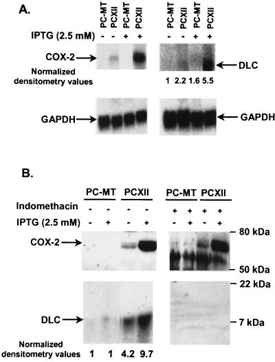
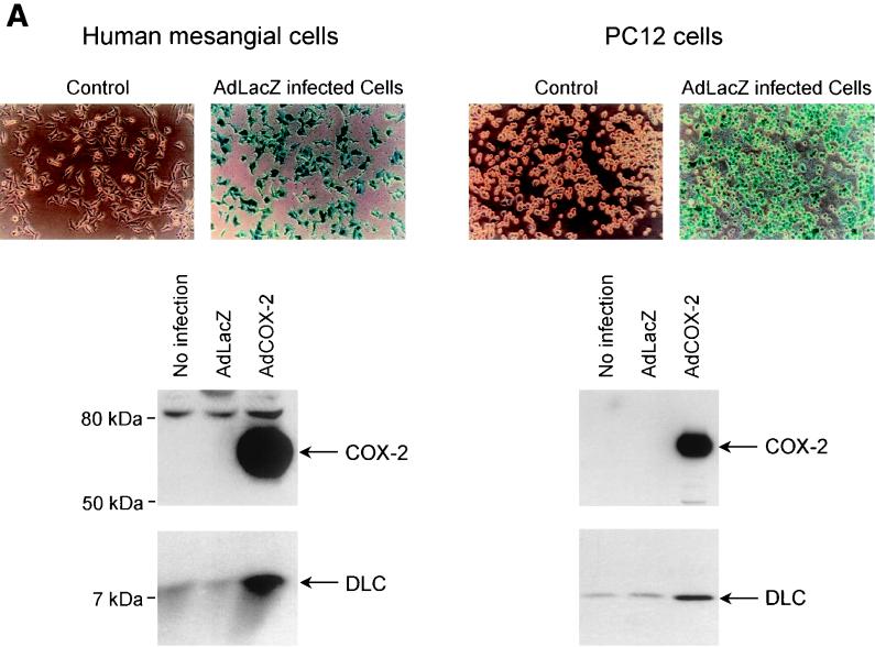
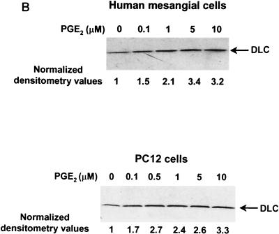
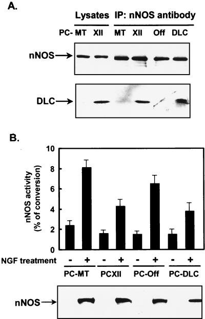
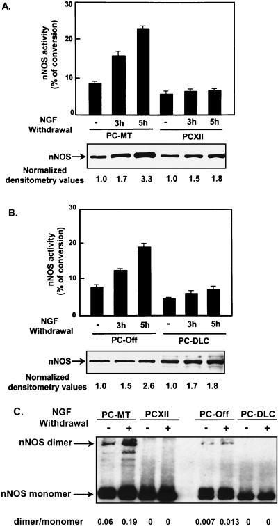
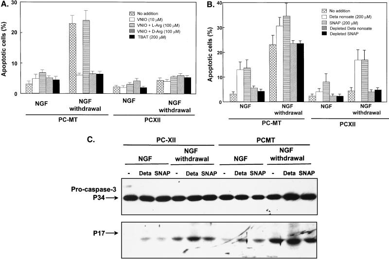
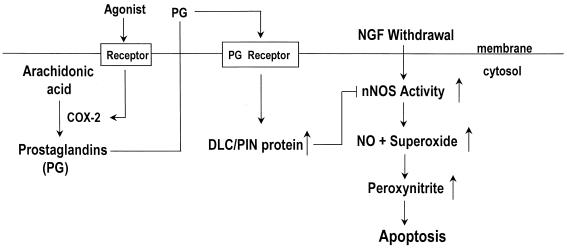
Cyclooxygenase 2 (COX-2) inhibits nerve growth factor (NGF) withdrawal apoptosis in differentiated PC12 cells. The inhibition of apoptosis by COX-2 was concomitant with prevention of caspase 3 activation. To understand how COX-2 prevents apoptosis, we used cDNA expression arrays to determine whether COX-2 regulates differential expression of apoptosis-related genes. The expression of dynein light chain (DLC) (also known as protein inhibitor of neuronal nitric oxide synthase [PIN]) was significantly stimulated in PC12 cells overexpressing COX-2. The COX-2-dependent stimulation of DLC expression was, at least in part, mediated by prostaglandin E(2). Overexpression of DLC also inhibited NGF withdrawal apoptosis in differentiated PC12 cells. Stimulation of DLC expression resulted in an increased association of DLC/PIN with neuronal nitric oxide synthase (nNOS), thereby reducing nNOS activity. Furthermore, nNOS expression and activity were significantly increased in differentiated PC12 cells after NGF withdrawal. This increased nNOS activity as well as increased nNOS dimer after NGF withdrawal were inhibited by COX-2 or DLC/PIN overexpression. An nNOS inhibitor or a membrane-permeable superoxide dismutase (SOD) mimetic protected differentiated PC12 cells from NGF withdrawal apoptosis. In contrast, NO donors induced apoptosis in differentiated PC12 cells and potentiated apoptosis induced by NGF withdrawal. The protective effects of COX-2 on apoptosis induced by NGF withdrawal were also overcome by NO donors. These findings suggest that COX-2 promotes cell survival by a mechanism linking increased expression of prosurvival genes coupled to inhibition of NO- and superoxide-mediated apoptosis.


Figures

FIG. 1
FIG. 1
COX-2 overexpression suppressed activation of caspase 3 after NGF withdrawal. PC-MT and PCXII cells were differentiated by NGF for 7 days in the presence of IPTG (2.5 mM), followed by NGF withdrawal for 0, 3, 6, 17, and 24 h. Cell lysates (40 μg) were separated by SDS–8 to 15% PAGE, and activation of caspase 3 was analyzed by Western blotting using caspase 3 antibody.
FIG. 2
FIG. 2
COX-2 overexpression in PC12 cells stimulated DLC expression. (A) Northern blot analysis of RNA from PC-MT and PCXII cells untreated or treated with IPTG for 17 h. Equal loading of RNA samples (10 μg) was confirmed by reprobing with GAPDH cDNA. Arrows indicate the positions of COX-2 and DLC. (B) Western blot analysis of cell lysates from PC-MT and PCXII cells untreated or treated with IPTG for 17 h. For indomethacin treatment, PC-MT and PCXII cells were pretreated with 10 μM indomethacin for 24 h and then incubated with or without IPTG for an additional 17 h. Cell lysates (30 μg) were resolved by SDS–8 to 20% PAGE, transferred onto a nitrocellulose membrane (0.22-μm pore size), and analyzed by Western blotting using COX-2 or DLC antibody. The densitometric values of the signals are normalized such that the zero time point is defined as 1.
FIG. 3
FIG. 3
Stimulation of DLC expression by transient expression of COX-2 using adenovirus gene delivery as well as by PGE2 treatment. (A) Human glomerular mesangial and PC12 cells were mock infected or infected with AdLacZ or AdCOX-2. As assessed by X-Gal staining, AdLacZ infected ∼100% of cells. Cell lysates (40 μg) were analyzed by Western blotting using anti-COX-2 or anti-DLC/PIN antibody. (B) Human mesangial cells were treated with increasing concentrations of PGE2, and control cells were treated with vehicle (dimethyl sulfoxide) for 6 h. Cell lysates (20 μg) from human mesangial and PC12 cells were separated by SDS–4 to 20% PAGE and analyzed by Western blotting using DLC/PIN antibody. Experiments were repeated three times with similar results.
FIG. 3
FIG. 3
Stimulation of DLC expression by transient expression of COX-2 using adenovirus gene delivery as well as by PGE2 treatment. (A) Human glomerular mesangial and PC12 cells were mock infected or infected with AdLacZ or AdCOX-2. As assessed by X-Gal staining, AdLacZ infected ∼100% of cells. Cell lysates (40 μg) were analyzed by Western blotting using anti-COX-2 or anti-DLC/PIN antibody. (B) Human mesangial cells were treated with increasing concentrations of PGE2, and control cells were treated with vehicle (dimethyl sulfoxide) for 6 h. Cell lysates (20 μg) from human mesangial and PC12 cells were separated by SDS–4 to 20% PAGE and analyzed by Western blotting using DLC/PIN antibody. Experiments were repeated three times with similar results.
FIG. 4
FIG. 4
Effects of DLC overexpression on differentiated PC12 cell apoptosis induced by NGF withdrawal. (A) Western blot analysis of PC12 cells stably expressing DLC. Lanes 1 and 2, PC-Off (parental) cells treated without (lane 1) or with (lane 2) doxycycline (10 μg/ml) for 48 h; lanes 3 and 4, PC-DLC cells stimulated without (lane 3) or with (lane 4) doxycycline (10 μg/ml) for 48 h. (B) Prevention of PC12 cell apoptosis induced by NGF withdrawal by DLC overexpression as determined by Hoechst staining. PC-Off and PC-DLC cells were differentiated by NGF for 7 days, followed by treatment with or without NGF withdrawal for 17 h. Data shown are the averages of three independent experiments, and error limits are within 3%. (C) Prevention of PC12 cell apoptosis induced by NGF withdrawal by DLC overexpression as determined by DNA fragmentation. Soluble cytoplasmic DNA from differentiated PC-Off or PC-DLC cells with (+) or without (−) NGF withdrawal for 17 h was analyzed by agarose gel electrophoresis. Some undegraded RNA was also strongly stained with ethidium bromide at the bottom of the gel. (D) Inhibition of caspase 3 activation induced by NGF withdrawal by DLC overexpression. Differentiated PC-Off or PC-DLC cells were incubated with or without anti-NGF antibody. At indicated time points, cells lysates (40 μg) were analyzed by Western blotting for caspase 3.
FIG. 5
FIG. 5
Association of DLC with nNOS in differentiated PC12 cells reduced nNOS activity. (A) PC-MT, PCXII, PC-Off, and PC-DLC cells were treated with NGF for 7 days; IPTG was added to PC12-MT and PCXII cells. Cell lysates (200 μg) were immunoprecipitated with nNOS antibody. Lysates (40 μg) and immunoprecipitates were analyzed by Western blotting using nNOS (top) or DLC (bottom) antibody. (B) nNOS activity was assayed in PC-MT, PCXII, PC-Off, and PC-DLC cells treated with or without NGF for 7 d; IPTG was added to PC-MT and PCXII cells. The activity of nNOS is presented as the percentage of conversion of [3H]arginine to [3H]citrulline. Values are means ± SD of three independent experiments. In the parallel experiments, cell lysates were analyzed for nNOS expression by Western blotting using nNOS antibody.
FIG. 6
FIG. 6
NGF withdrawal from differentiated PC12 cells further stimulated nNOS expression and activity, and nNOS activity was inhibited by COX-2 or DLC overexpression. (A and B) PC-MT, PCXII, PC-Off, and PC-DLC cells were treated with NGF for 7 days; IPTG was added to PC-MT and PCXII cells. At 0, 3, and 5 h after NGF withdrawal, nNOS expression was analyzed by Western blotting, and activity was measured as described in Materials and Methods. Values are means ± SD of three experiments. (C) Cell lysates from differentiated PC-MT, PCXII, PC-Off, or PC-DLC cells incubated with (+) or without (−) anti-NGF antibody for 6 h were analyzed for nNOS dimerization by SDS-PAGE under low-temperature conditions as described previously (31), followed by Western blot analysis using nNOS antibody. The dimer/monomer ratio was determined by densitometrically scanning of both nNOS bands on autoradiogram.
FIG. 7
FIG. 7
Effects of NO donors, nNOS inhibitor, or manganese TBAT on apoptosis in differentiated PC12 cells. (A) Differentiated PC-MT and PCXII cells were treated with or without nNOS inhibitor as indicated in the presence of NGF (25 ng/ml) for 24 h or anti-NGF antibody for 17 h. Nuclei were stained with Hoechst 33528. Cells containing condensed or fragmented nuclei were scored as apoptotic. (B) Differentiated PC-MT and PCXII cells were treated with or without NO donor as indicated in the presence of NGF (25 ng/ml) for 24 h or anti-NGF antibody (1:1,000 dilution) for 17 h. For depletion, the NO donors were incubated at room temperature in serum-free medium for 4 days to completely liberate NO. Values are means ± SD of four experiments. (C) Differentiated PC-MT or PCXII cells, treated with or without NO donor (200 μM DETA-NONOate [Deta] or SNAP) in the presence of NGF (25 ng/ml) or anti-NGF antibody (1:500 dilution) for 6 h. Cell lysates (40 μg) were separated by SDS–8 to 15% PAGE, and activation of caspase 3 was analyzed by Western blotting using caspase 3 antibody.
FIG. 8
FIG. 8
Model for regulation of apoptosis by COX-2. Removal of NGF from differentiated PC12 cells elevates nNOS expression and activity, thereby leading to production of excess NO and superoxide, which contributes to cell death by producing peroxynitrite. Stimulation of COX-2 expression by agonists is concomitant with increased production of PGE2. PGE2 is released from the cells and can stimulate the prostaglandin (PG) receptors on PC12 cells, leading to an increase in DLC expression. Stimulation of DLC expression results in an increase in association of DLC with nNOS, causing inactivation of nNOS and thus reducing production of NO and superoxide. This may prevent PC12 cells apoptosis induced by NGF withdrawal.

References

    1. Albina J E, Cui S, Mateo R B, Reichner J S. Nitric oxide-mediated apoptosis in murine peritoneal macrophages. J Immunol. 1993;150:5080–5085. - PubMed
    1. Babu B R, Griffith O W. N5-(1-Imino-3-butenyl)-l-ornithine. A neuronal isoform selective mechanism-based inactivation of nitric oxide synthase. J Biol Chem. 1998;273:8882–8889. - PubMed
    1. Becker A J, Gillardon F, Blumcke I, Langendorfer D, Beck H, Wiestler O D. Differential regulation of apoptosis-related genes in resistant and vulnerable subfields of the rat epileptic hippocampus. Mol Brain Res. 1999;67:172–176. - PubMed
    1. Benashski S E, Harrison A, Patel-King R S, King S M. Dimerization of highly conserved light chain shared by dynein and myosin V. J Biol Chem. 1997;272:20929–20935. - PubMed
    1. Chammugam P, Feng L, Liou S, Jang B C, Boudreau M, Yu G, Le J H, Kwon H J, Beppu T, Yoshida M, Xia Y, Wilson C B, Hwang D. Radicicol, a protein tyrosine kinase inhibitor, suppresses the expression of mitogen-inducible cyclooxygenase in macrophages stimulated with lipopolysaccharide and in experimental glomerulonephritis. J Biol Chem. 1995;270:5418–5426. - PubMed

Publication types

MeSH terms

Substances