. 2000 Nov;20(22):8602-12.
doi: 10.1128/MCB.20.22.8602-8612.2000.

Chromatin association of human origin recognition complex, cdc6, and minichromosome maintenance proteins during the cell cycle: assembly of prereplication complexes in late mitosis

Affiliations

Chromatin association of human origin recognition complex, cdc6, and minichromosome maintenance proteins during the cell cycle: assembly of prereplication complexes in late mitosis

J Méndez et al. Mol Cell Biol. 2000 Nov.

Abstract

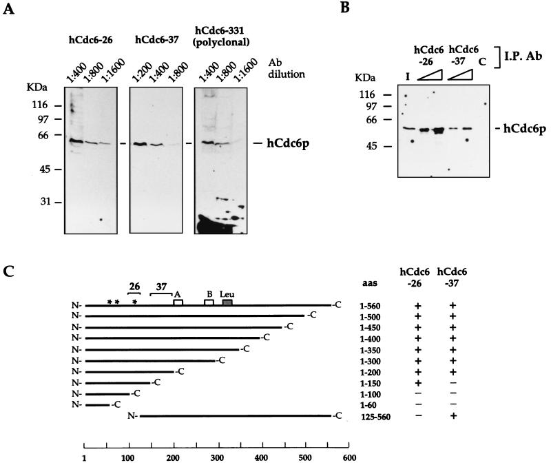
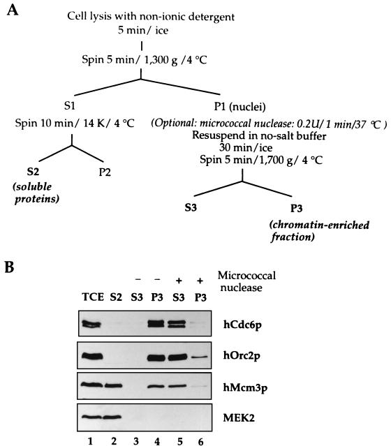
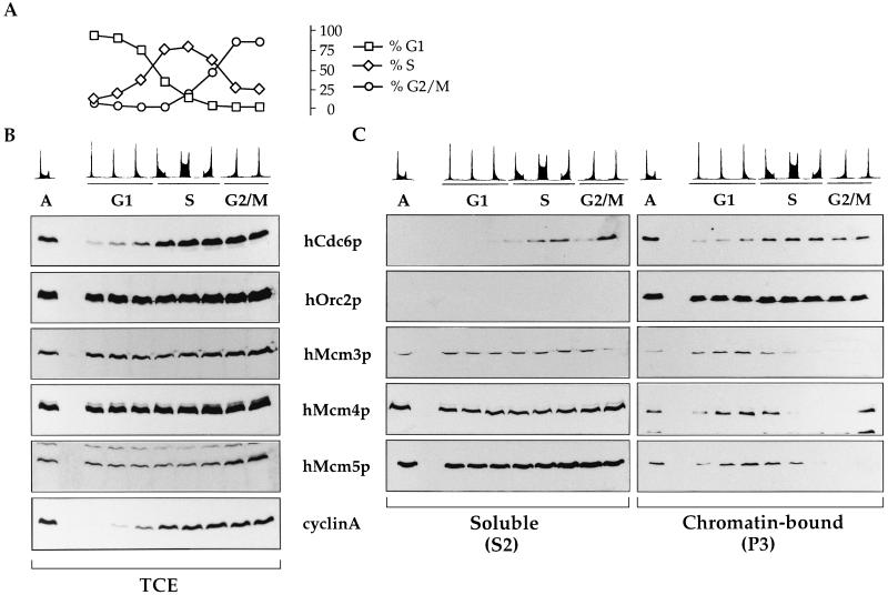
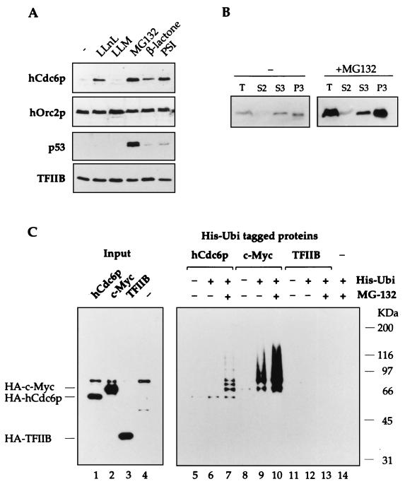
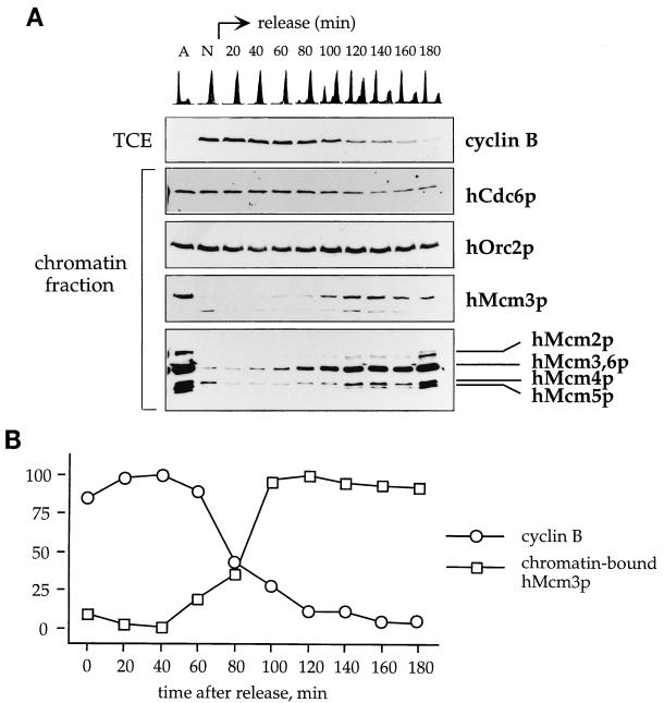
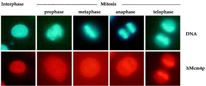
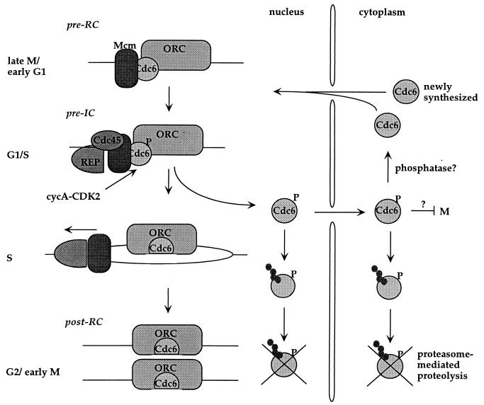
Evidence obtained from studies with yeast and Xenopus indicate that the initiation of DNA replication is a multistep process. The origin recognition complex (ORC), Cdc6p, and minichromosome maintenance (MCM) proteins are required for establishing prereplication complexes, upon which initiation is triggered by the activation of cyclin-dependent kinases and the Dbf4p-dependent kinase Cdc7p. The identification of human homologues of these replication proteins allows investigation of S-phase regulation in mammalian cells. Using centrifugal elutriation of several human cell lines, we demonstrate that whereas human Orc2 (hOrc2p) and hMcm proteins are present throughout the cell cycle, hCdc6p levels vary, being very low in early G(1) and accumulating until cells enter mitosis. hCdc6p can be polyubiquitinated in vivo, and it is stabilized by proteasome inhibitors. Similar to the case for hOrc2p, a significant fraction of hCdc6p is present on chromatin throughout the cell cycle, whereas hMcm proteins alternate between soluble and chromatin-bound forms. Loading of hMcm proteins onto chromatin occurs in late mitosis concomitant with the destruction of cyclin B, indicating that the mitotic kinase activity inhibits prereplication complex formation in human cells.


Figures

FIG. 1
FIG. 1
Specificities of new monoclonal anti-hCdc6p antibodies (Ab). (A) Nuclear extracts from asynchronous 293 cells were subjected to SDS-PAGE, and proteins were transferred to nitrocellulose. After protein staining with Ponceau-S red, individual lanes were cut and immunoblotted with the indicated dilution of monoclonal antibody hCdc6-26 or hCdc6-37 or a polyclonal antibody, anti-hCdc6p. A major signal corresponding to a 62-kDa protein was detected in all cases. (B) Immunoprecipitation (I.P.). Fifty microliters of nuclear extract from 293 cells (0.5 mg of total protein) was incubated for 1 h with 1 or 3 μl of ascitic fluid of the corresponding monoclonal antibody or with 3 μl of an unrelated control antibody (lane C). Immunocomplexes were purified with protein G-Sepharose 4B, subjected to SDS-PAGE, transferred to nitrocellulose, and immunoblotted with anti-hCdc6p polyclonal antibodies. Lane I, 10% of the input sample. (C) Epitope mapping. Constructs expressing different C-terminal deletions and one N-terminal deletion of hCdc6p were made and expressed in E. coli as GST fusion proteins. Monoclonal antibodies hCdc6-26 and hCdc6-37 were used to detect the different truncated proteins by immunoblotting. The asterisks represent consensus sites for CDK phosphorylation. Boxes A and B indicate the position of Walker A and Walker B motifs, required for ATP binding and/or hydrolysis. The gray box marked Leu represents the hCdc6p leucine zipper. The regions that contain the epitopes recognized by hCdc6-26 and hCdc6-37 are indicated with brackets. aa, amino acid.
FIG. 2
FIG. 2
Chromatin binding of initiation proteins. (A) Scheme of the biochemical fractionation method. See Materials and Methods for details. (B) An asynchronous culture of Raji cells was subjected to the biochemical fractionation described in panel A. After cell lysis, the nuclei were divided in two aliquots. One of them was incubated for 1 min at 37°C with 0.2 U of micrococcal nuclease, and the other one was incubated in the same conditions without nuclease. After this treatment, nuclei were lysed and the solubilized nuclear proteins (S3) were separated from the chromatin-bound proteins (P3) by centrifugation. The distributions of different proteins in the total cell extract (TCE) soluble fraction (S2), solubilized nuclear proteins fraction (S3), and chromatin-nuclear matrix-bound fraction (P3) are shown.
FIG. 3
FIG. 3
Protein levels and chromatin association of hCdc6p, hOrc2p, and hMcm proteins during the cell cycle. An asynchronous culture of human Raji cells was subjected to centrifugal elutriation to isolate cells at different points of the cell cycle. The DNA content for the cells in each fraction is shown. (A) Percentage of cells in each phase of the cell cycle, estimated with the CellFIT computer program. (B) Levels of initiator factors in total cell extracts (TCE). Equivalent amounts of each total cell extract (normalized by cell number) were subjected to SDS-PAGE and transferred to nitrocellulose for immunobloting with the indicated antibodies. The concentration of cyclin A is also shown as a control of cell cycle progression. Lane A, asynchronous cells. (C) Cell cycle-regulated chromatin association of hOrc2p, hCdc6p, and hMcm proteins. Cells at different points in the cell cycle were subjected to the biochemical fractionation described for Fig. 2A. Immunoblots of the soluble protein fraction (S2) or chromatin-enriched fraction (P3) are shown.
FIG. 4
FIG. 4
hCdc6p is targeted by ubiquitination for destruction by the proteasome. (A) Stabilization of hCdc6p by inhibitors of the proteasome. After treatment of HeLa cells with the indicated inhibitor, the cells were harvested and used to prepare total cell extracts. The steady-state levels of hCdc6p, as well as p53, increased in the presence of proteasome inhibitors but not in the presence of calpain inhibitor LLM. The levels of hOrc2p or TFIIB did not change significantly. (B) HeLa cells treated with dimethyl sulfoxide (control) or MG132 were subjected to the biochemical fractionation described for Fig. 2A. The levels of hCdc6p in the different fractions are shown. (C) In vivo polyubiquitination of hCdc6p. HeLa cells were transfected with plasmids expressing HA-tagged versions of hCdc6p, c-Myc, or TFIIB in the absence or in the presence of a plasmid that expresses His-Ubi. When indicated, 20 μM MG132 was added at 24 h posttransfection. Cells were harvested and lysed at 36 h after transfection. His-tagged proteins were purified and subjected to SDS-PAGE. After transfer to nitrocellulose, the samples were immunoblotted with anti-HA antibodies. Lanes 1 to 4 show 6% of the input protein.
FIG. 5
FIG. 5
Chromatin association of hMcm proteins at the M/G1 transition. (A) HeLa cells were synchronized at early mitosis with nocodazole (see Materials and Methods), and a fraction of cells was collected at different time points after release from the block. An aliquot of the cells isolated at each time point was used to determine the DNA content by flow cytometry (top panel), and the rest were subjected to the biochemical fractionation described for Fig. 4A. The presence of hCdc6p, hOrc2p, and hMcm proteins in the chromatin-enriched fraction (P3) was analyzed. The top panel shows the progressive degradation of cyclin B in total cell extracts (TCE) as the cells progress through mitosis. Lane A, asynchronous culture; lane N, nocodazole-arrested cells. (B) Quantitation of total cyclin B and chromatin-bound hMcm3p in the experiment shown in panel A. The results are expressed as percentages of the maximum signal in each curve.
FIG. 6
FIG. 6
hMcm4p is loaded onto chromatin during late mitosis and early G1. The subcellular localization of hMcm4p was addressed by indirect immunofluorescence in HeLa cells. Cells were identified as interphasic or mitotic by direct observation of the chromatin condensation state, and the different stages of mitosis were determined by observation of the chromosome distribution. Representative photographs of each stage are shown. hMcm4p stains the nuclei of interphasic cells and the whole cell in prophase, metaphase, or anaphase. In the latter two phases, exclusion of staining in the chromatin is observed. In contrast, strong hMcm4p staining is detected in the chromatin of late telophase cells, which have not yet completed cytokinesis.
FIG. 7
FIG. 7
A model for the regulation of hCdc6p in the cell cycle. See the text for details. IC, initiation complex.

References

    1. Aparicio O M, Weinstein D M, Bell S P. Components and dynamics of DNA replication complexes in S. cerevisiae: redistribution of MCM proteins and Cdc45p during S phase. Cell. 1997;91:59–69. - PubMed
    1. Bell S P, Stillman B. ATP-dependent recognition of eukaryotic origins of DNA replication by a multiprotein complex. Nature. 1992;357:128–134. - PubMed
    1. Bolt M W, Mahoney P A. High-efficiency blotting of proteins of diverse sizes following sodium dodecyl sulfate-polyacrylamide gel electrophoresis. Anal Biochem. 1997;247:185–192. - PubMed
    1. Bueno A, Russell P. Dual functions of CDC6: a yeast protein required for DNA replication also inhibits nuclear division. EMBO J. 1992;11:2167–2176. - PMC - PubMed
    1. Chong J P, Hayashi M K, Simon M N, Xu R M, Stillman B. A double-hexamer archaeal minichromosome maintenance protein is an ATP-dependent DNA helicase. Proc Natl Acad Sci USA. 2000;97:1530–1535. - PMC - PubMed

Publication types

MeSH terms

Substances