. 2000 Oct;5(4):291-305.
doi: 10.1379/1466-1268(2000)005<0291:tdohsp>2.0.co;2.

The distribution of heat shock proteins in the nervous system of the unstressed mouse embryo suggests a role in neuronal and non-neuronal differentiation

Affiliations

The distribution of heat shock proteins in the nervous system of the unstressed mouse embryo suggests a role in neuronal and non-neuronal differentiation

M T Loones et al. Cell Stress Chaperones. 2000 Oct.

Abstract

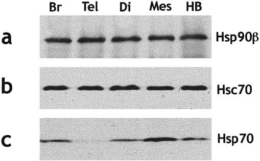
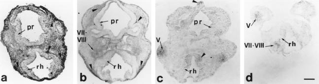
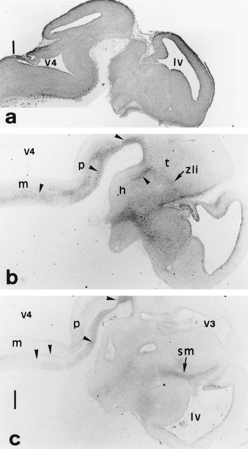
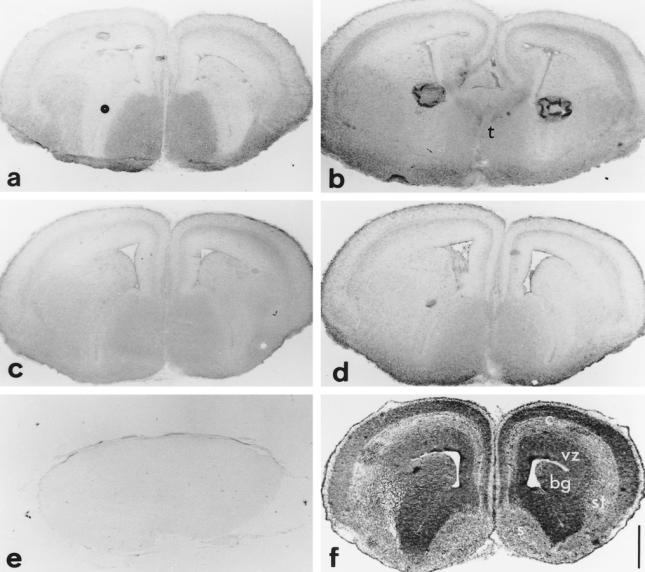
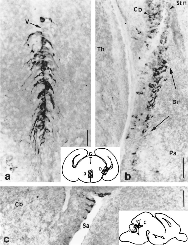
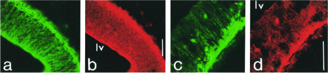
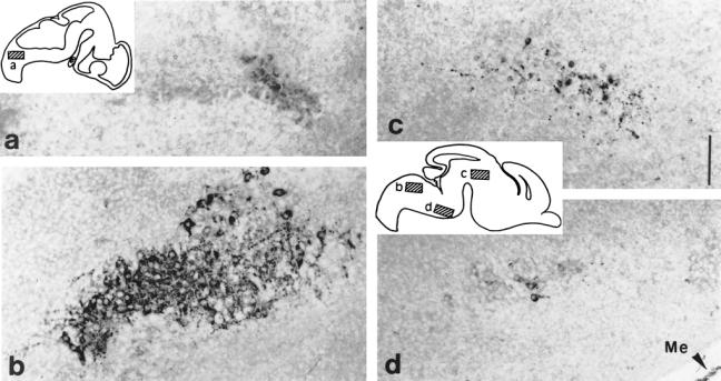
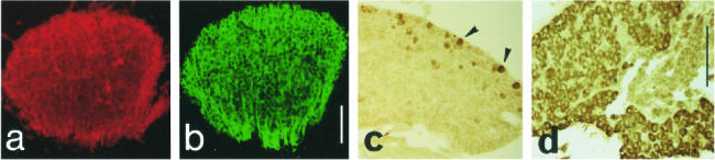
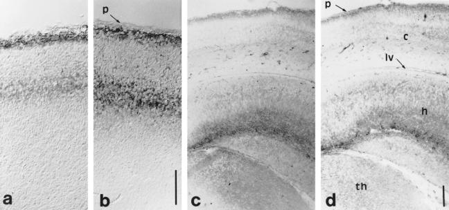
Heat shock proteins (Hsps) act as molecular chaperones and are generally constitutively expressed in the absence of stress. Hsps are also inducible by a variety of stressors whose effects could be disastrous on the brain. It has been shown previously that Hsps are differentially expressed in glial and neuronal cells, as well as in the different structures of the brain. This differential expression has been related to specific functions distinct from their general chaperone function, such as intracellular transport. We investigated here the constitutive expression of 5 Hsps (the small Hsp, Hsp25, the constitutive Hsc70 and Hsp90beta, the mainly inducible Hsp70 and Hsp90alpha), and of a molecular chaperone, TCP-1alpha during mouse nervous system development. We analyzed, by immunohistochemistry, their distribution in the central nervous system and in the ganglia of the peripheral nervous system from day 9.5 (E9.5) to day 17.5 (E17.5) of gestation. Hsps are expressed in different cell classes (neuronal, glial, and vascular). The different proteins display different but often overlapping patterns of expression in different regions of the developing nervous system, suggesting unique roles at different stages of neural maturation. Their putative function in cell remodeling during migration or differentiation and in protein transport is discussed. Moreover we consider Hsp90 function in cell signaling and the role of Hsp25 in apoptosis protection.


Figures

Fig 1.
Fig 1.
Comparative Western blot analysis of Hsps level in the brain of E16.5 mouse embryos. Samples containing 20 μg of proteins were loaded per lane for whole brain (Br), telencephalon (Tel), diencephalon (Di), mesencephalon (Mes), and hindbrain (HB). Hsp90β (a), Hsc70 (b), and Hsp70 (c) were successively analyzed. Hsp90β and Hsc70 show comparable levels in each brain region, whereas the amount of Hsp70 is greater in mes- than in di-, telencephalon, or hindbrain
Fig 2.
Fig 2.
Hsp detection on cephalic coronal sections of an E9.5 mouse embryo. Sections cut the neural tube twice, at the prosencephalon (pr) and at the rhombencephalon (rh) levels. (a) Hsp90β ubiquitous is enriched along the pathway of the neural crest cells (NCC) and ganglion condensations (arrowheads); (b) Hsp25: migration paths of the NCC in the cephalic mesenchyme (arrowheads) and condensations of the VII–VIII ganglia (arrow) are enriched in Hsp25. (c) Hsc70: tectum of the forebrain, basal plate of the medulla (arrowheads), and the protrusion of the trigeminal (V) ganglion (arrow) are revealed by the antibody. (d) TCP-1α is localized on trigeminal (V) and on acoustico-facial (VII–VIII) condensations (arrows). Scale 100 μm
Fig 3.
Fig 3.
(a) Hsp90β, (b) Hsp25, and (c) Hsc70 distribution on E12.5 neural tube parasagittal sections. (b,c) Focused at the forebrain and the ventral hindbrain levels. (a) Hsp90β displays an ubiquitous distribution in the marginal zone. (b,c) The longitudinal tracts along the pons (p) and the medulla (m) (arrowheads), the zona limitans interthalamica (zli), a bundle that limits prosomeres in the diencephalon and the peripheral descending tracts, as the stria medullaris (sm) are recognized by anti-Hsp25 and anti-Hsc70. h, Hypothalamus; lv, lateral ventricle of telencephalon; t, thalamus; v3, v4, third and fourth ventricles. Scale 250 μm
Fig 4.
Fig 4.
Hsc70 (a), Hsp25 (b), Hsp90α (c) and Hsp90β (d) distribution on E15.5 telencephalic coronal sections compared to toluidine blue staining (f) and to a negative control section where the first antibody was omitted (e). Hsc70, Hsp25, and the Hsp90s are differentially expressed in some layers of the developing stratification of the cortex (c), in septum (s), basal ganglia (bg), striatum (st), but not in the ventricular zone (vz). The more caudal section (b) shows that Hsp25 is also expressed in thalamic (t) structures. Note in (b) that the strong staining in the middle of each hemisphere is an artifact due to a bubble of nonadherant material. Toluidine blue intensely (f) stains the differentiating cortex (c) and the ventricular zone (vz), regions where the cell nuclei population is very dense. Scale 500 μm.
Fig 5.
Fig 5.
Hsp90α exhibits a highly specific distribution on the E17.5 brain hypothalamic coronal sections (a,b) and mes-metencephalic parasagittal section (c). Schematic drawings of these sections localize the high power pictures (a–c). (a) Hypothalamic neurons of the putative dorsomedial nuclei on both sides of the obturated third ventricle (V). (b) Neurons of the bed nucleus of the stria terminalis (Bn) are strongly labeled as are the choroid plexus (Cp) of the lateral ventricle to a lesser extent. pa, Pallidum; stn, strionuclear neuroepithelium; Th, thalamus. (c) Few neurons of the neuroepithelium along the border of the Sylvius aqueduct (Sa) of the isthmus (mes-metencephalic limit) (cb) cerebellum. Scales 100 μm
Fig 6.
Fig 6.
Hsp25 double labeling on E12.5 cortical parasagittal sections with, respectively, RC2 and TuJ1. (a) RC2 recognizes the early differentiating radial glial cells in the ventricular zone, and (b) Hsp25 displays a partially coincident localization with RC2. (c) TuJ1 strongly labels the early differentiating cortical neurons of the cortical plate, and (d) Hsp25 labels similarly. lv, Lateral ventricle. Scales 100 μm
Fig 7.
Fig 7.
Hsp25 expression in brain nuclei: E12.5 and E15.5 parasagittal sections. Diagrams of these sections localize the level of highpower pictures: E12.5 (a), E15.5 (b–d) (a) Nascent hypoglossal neurons start to express Hsp25. (b) Cytoplasmic presence of Hsp25 in the somata and processes of the large motoneurons of the hypoglossal nucleus at E15.5. (c) Neurons of an indeterminate nucleus in the differentiating field of the tegmentum. (d) Some neurons of the ventral area of the pontic region express Hsp25. Notice that among the meningae (Me), the dura mater is strongly labeled with anti-Hsp25. Scale 250 μm.
Fig 8.
Fig 8.
Hsp25 (a,c) and Hsp90α (d) during PNS ganglia differentiation. (a) E12.5 coronal section: Hsp25 is highly present in all differentiating neurons of the trigeminal ganglion. (b) The same neurons are labeled with TuJ1. (c) E15.5 parasagittal section: only a subpopulation of the neurons of the trigeminal ganglion is labeled with anti-Hsp25 (arrowheads). (d) E17.5 coronal section: most neurons express Hsp90α. Scales 150 μm
Fig 9.
Fig 9.
Hsp70 localization on a E15.5 brain parasagittal section. In the forebrain derivates Hsp70 is present in the external layer of the olfactory bulb (ob), the cortex (c) (to a lesser extent in hippocampus [h]), in the septum, the preoptic area (po), and the fibers of the diencephalic tracts. On this section the labeled stria medullaris corresponds with the area of the posterior commissure (pc) and the labeling for the mammilothalamic tract (mt) corresponds with the caudal area of the mammilar region. In the midbrain Hsp70 is present in the inferior colliculus (ic) and in the ventral tegmental nucleus (vt). In the caudalmost region of the brain the immunolabeling is distributed in the central vermix of the cerebellum (cb), in the prepositus (pp), and in the cuneate (cu) nuclei of the dorsal medulla (m). p, Pons. Scale 500 μm
Fig 10.
Fig 10.
Hsp70 (a,c) and GFAP (b,d) localization on E15.5 (a,b) and E17.5 (c,d), respectively, parasagittal and coronal sections of the cortex. (a,b) Hsp70 (a) is differentially and specifically expressed throughout 2 of the 6 layers of the newly organized stratification of the neocortex as GFAP (b). (c,d) Hsp70 (c) is heavily expressed under the pial surface and in the hippocampus (h) as is GFAP (d). c, neocortex; h, hippocampus; lv, lateral ventricle; p, pial surface; th, thalamus. Scales 100 μm

Similar articles

Cited by

References

    1. Ali A, Bharadwaj S, O'Carroll R, Ovsenek N. HSP90 interacts with and regulates the activity of heat shock factor 1 in Xenopus oocytes. Mol Cell Biol. 1998;18:4949–4960. - PMC - PubMed
    1. Altman J, Bayer SA. Development of the brain stem in the rat. I. Thymidine-radiographic study of the time of origin of neurons of the lower medulla. J Comp Neurol. 1980;194:1–35. - PubMed
    1. Altman J, Bayer SA. The development of the rat hypothalamus. Adv Anat Embryol Cell Biol. 1986;100:1–178. - PubMed
    1. Altman J, Bayer SA 1995 Atlas of Prenatal Rat Brain Development. CRC Press, Boca Ràton, FL.
    1. Aquino DA, Klipfel AA, Brosnan CF, Norton WT. The 70-kDa heat shock cognate protein (HSC70) is a major constituent of the central nervous system and is up-regulated only at the mRNA level in acute experimental autoimmune encephalomyelitis. J Neurochem. 1993;61:1340–1348. - PubMed

Publication types

MeSH terms

LinkOut - more resources