. 2000 Nov;84(11):1269-74.
doi: 10.1136/bjo.84.11.1269.

Clinicopathological correlation of deep retinal vascular anomalous complex in age related macular degeneration

Affiliations

Clinicopathological correlation of deep retinal vascular anomalous complex in age related macular degeneration

B A Lafaut et al. Br J Ophthalmol. 2000 Nov.

Abstract

Aims: To analyse the histopathology of "deep retinal vascular anomalous complex" or "chorioretinal anastomosis".

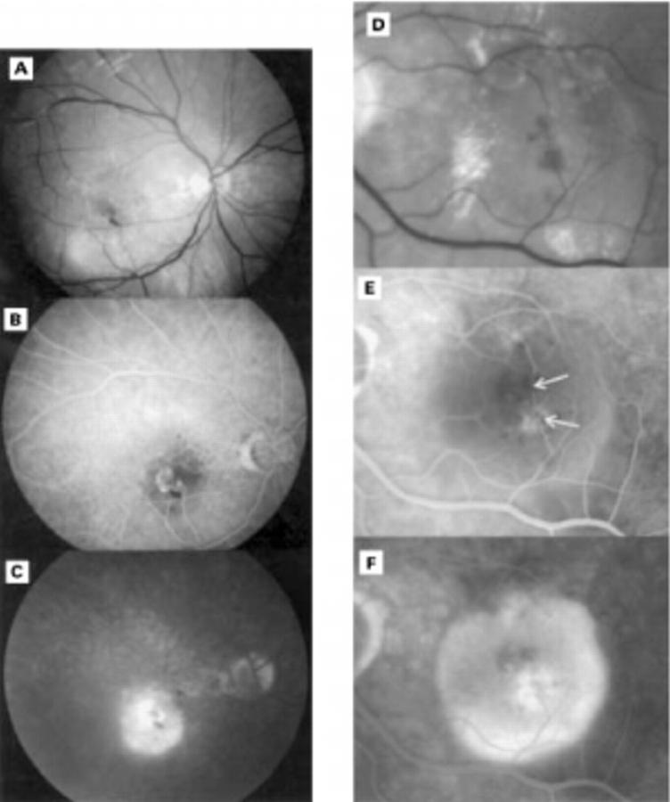
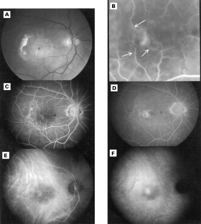
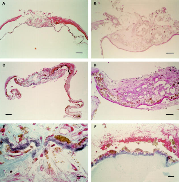
Methods: Six patients with a deep retinal vascular anomalous complex (age range 66-88 years) had fundus photography and fluorescein angiography not more than 14 days before foveal translocation surgery. Four patients were also documented with indocyanine green angiography. The surgical specimens were serially sectioned and stained in a stepped fashion with Masson trichrome, periodic acid Schiff, and phosphotungstic acid haematoxylin, a histochemical stain for fibrin.

Results: A subretinal fibrovascular membrane was surrounded by a rim consisting of diffuse drusen (basal laminar deposits), retinal pigment epithelium, and amorphous, fibrinous material interspersed with remains of outer segments in all specimens. In two specimens vascular structures were identified that left the specimen towards the retina. Amorphous material with the remains of outer segments was not found on the retinal side of the fibrovascular tissue itself but in four specimens a small neuroretinal portion (outer nuclear layer) was adherent to the complex. In three specimens a thin fibrocellular membrane was seen at the choroidal side of the diffuse drusen.

Conclusion: Deep retinal vascular anomalous complex represents histologically neovascularisation growing out of the neuroretina, into the subretinal space, which mimics choroidal neovascularisation. The term therefore appears rightly chosen.


Figures

Figure 1
Figure 1
(A), (B), and (C) respectively red-free, venous, and late phase fluorescein angiography of a 71 year old patient. Intraretinal lipid exudates and haemorrhages are seen on the red-free photograph. A subretinal neovascular net is seen in the right eye which appears connected to at least two retinal vessels in the early phase. The hyperfluorescence of this deep retinal vascular anomalous complex persists in the late phase. The associated retinal pigment epithelial detachment appears hypofluorescent in the early phase but becomes hyperfluorescent in the late phase. (D), (E), and (F) respectively enlarged red-free, venous, and late phase fluorescein angiography of a 70 year old patient. Intraretinal lipid exudates and haemorrhages are seen, arrows indicate retinal vessels that appear to dip into the deep retinal vascular anomalous complex. The associated serous pigment epithelial detachment is clearly demonstrated in the late phase.
Figure 2
Figure 2
Red-free (A) fluorescein angiogram (B, C, and D) and indocyanine green angiogram of a 73 year old patient. On the red-free photograph, intraretinal lipid exudates, haemorrhages, and a drusenoid pigment epithelial detachment can be seen. Fluorescein angiography indicates a deep retinal vascular anomalous complex that appears to be connected to three retinal vessels (arrows). The venous (E) and late phase (F) indocyanine green angiogram identifies a vascular net. The lesion persists as a hot spot in the late phase, entirely within a hypofluorescent zone corresponding to the retinal pigment epithelial detachment.
Figure 3
Figure 3
(A) MTC stain, bar = 100 µm, overview of specimen corresponding to Figure 2. A subretinal central fibrovascular nodule is seen surrounded by a rim composed of amorphous material, retinal pigment epithelium, and diffuse drusen. (B) PAS stain, bar = 50 µm, detail of same specimen as (A). The fibrovascular tissue immediately contacts the diffuse drusen. Some vessels leave the specimen towards the retina. (C) MTC stain, bar = 100 µm, overview of specimen corresponding to Figure 1A, B, C. A central fibrovascular nodule is seen, consisting both of a subretinal vascular component and an intra-Bruch's fibrocellular component, with surrounding rim. (D) PAS stain, bar = 50 µm, detail of same specimen as (C). A rich vasculature is demonstrated in the subretinal part of the specimen. (E) MTC stain, bar 10 µm, detail of same specimen as (C). An interruption of the diffuse drusen layer is seen with a fibrous tissue connecting the subretinal with the intra-Bruch's component. (F) MTC stain, bar 25 µm, detail of a third specimen, at the level of the rim. Pale staining, fine granular material is seen at the choroidal side of the diffuse drusen.

References

    1. Retina. 1999;19(4):332-5 - PubMed
    1. Postgrad Med J. 1998 Apr;74(870):203-7 - PubMed
    1. Arch Ophthalmol. 1971 Nov;86(5):487-94 - PubMed
    1. Invest Ophthalmol. 1973 Jan;12(1):65-76 - PubMed
    1. Br J Ophthalmol. 1973 Dec;57(12):951-65 - PubMed