. 2000 Nov 1;20(21):7863-70.
doi: 10.1523/JNEUROSCI.20-21-07863.2000.

Phorbol esters potentiate evoked and spontaneous release by different presynaptic mechanisms

Affiliations

Phorbol esters potentiate evoked and spontaneous release by different presynaptic mechanisms

J Waters et al. J Neurosci. .

Abstract

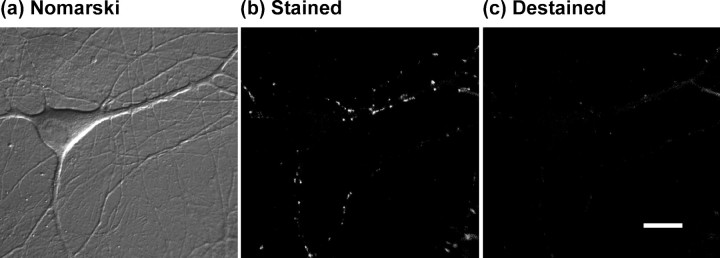
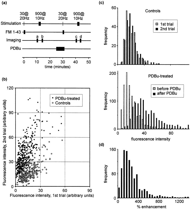
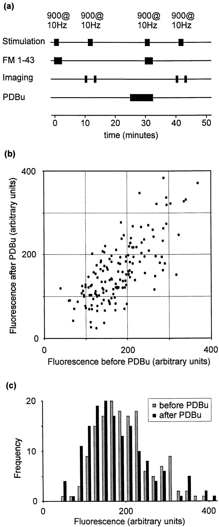
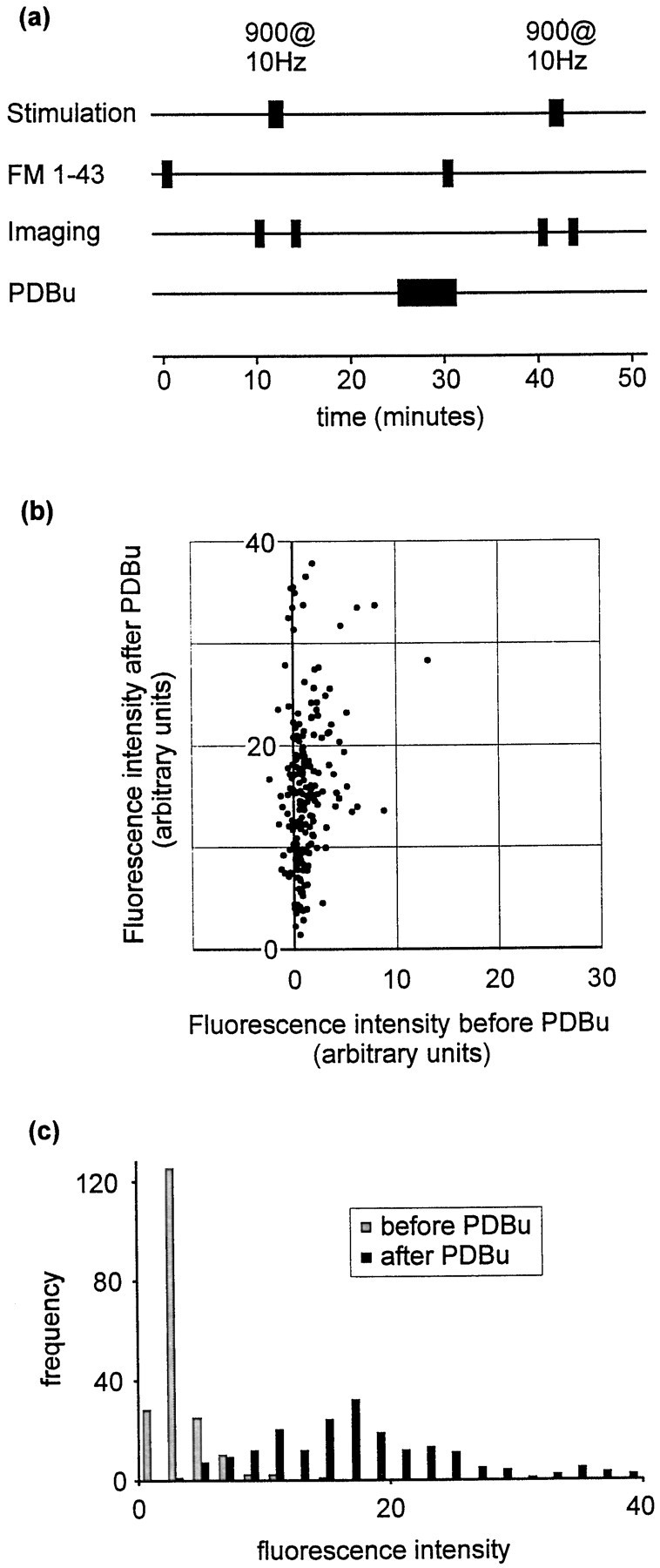
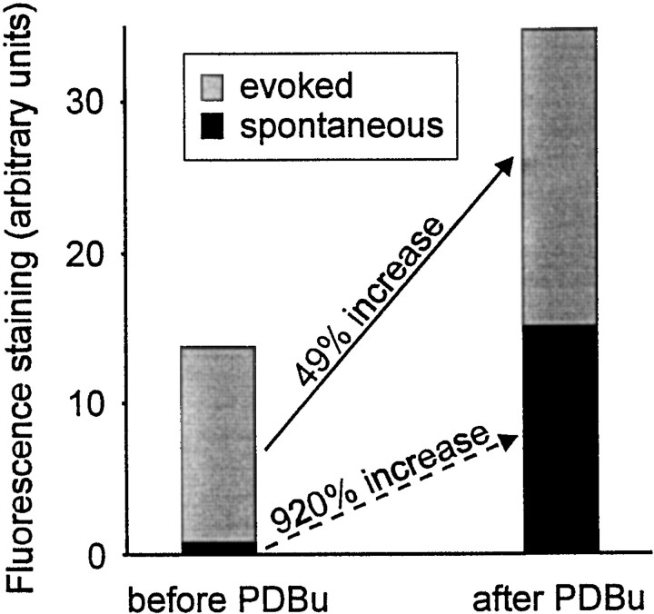
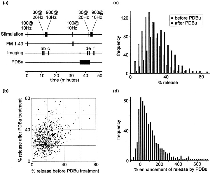
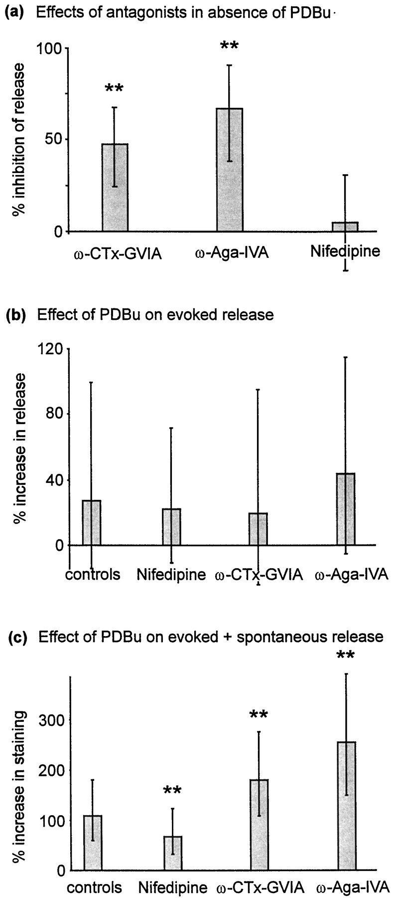
Phorbol esters enhance release from a variety of cell types. The mechanism by which phorbol esters potentiate presynaptic release from central neurons is unclear, although effects of phorbol esters both on the readily releasable pool of vesicles and on presynaptic calcium channels have been shown. Using confocal microscopy and the fluorescent styryl dye FM 1-43, we have examined the effects of phorbol-12,13-dibutyrate (PDBu) on presynaptic vesicle turnover at individually identified synapses in dissociated cultures obtained from neonatal rat hippocampus. Using different dye staining and destaining protocols we were able to resolve two effects of PDBu. Potentiation of evoked release by PDBu was insensitive to calcium channel antagonists, suggesting that this effect results from an increased number of vesicles in the readily releasable pool. Since we observed no effect of PDBu on the size of the total recycling vesicle pool, we conclude that phorbol esters alter the equilibrium between reserve and readily releasable pools. An additional effect of PDBu on spontaneous release was observed. This effect was antagonized by nifedipine but not omega-conotoxin GVIA or omega-agatoxin IVA. We conclude that PDBu influences spontaneous and evoked release by two different mechanisms: through L-type calcium channels and through an increase in the proportion of recycling vesicles in the readily releasable pool. In addition to further clarifying the mechanism of action of phorbol esters, these results suggest that phorbol esters may be a useful tool with which to probe the function of the readily releasable pool of presynaptic vesicles at CNS synapses.


Figures

Fig. 1.
Fig. 1.
Example of FM 1-43 staining.a, Nomarski image of a pyramidal neuron in a mixed neuronal–glial dissociated culture. b, Fluorescence image of the same field acquired after FM 1-43 staining, using a 100-stimulus train at 10 Hz and a 40 sec exposure to FM 1-43. Discrete fluorescent puncta are visible, often at sites corresponding to dendritic interactions visible in the Nomarski image. c, Fluorescence image acquired after destaining using a 900-stimulus train at 10 Hz. Note that the punctate staining is primarily absent, leaving a dim background attributable to nonspecific membranous staining by FM 1-43. Scale bar, 10 μm.
Fig. 2.
Fig. 2.
Effect of PDBu on staining.a, Summary of the staining–destaining protocol used. b, Scatter plot showing fluorescence intensities for individual fluorescent puncta in two consecutive trials. PDBu resulted in increased FM 1-43 staining (filled symbols), whereas controls (open symbols) displayed no mean change in staining. c, Data represented as frequency histograms. Top panel, control data; bottom panel, effect of PDBu treatment.d, Frequency histogram showing the percentage increase in staining observed after PDBu exposure.
Fig. 3.
Fig. 3.
PDBu and total recycling pool size.a, Schematic illustration of the protocol used to examine the effect of phorbol treatment on total recycling pool size.b, Scatter plot comparing fluorescence staining before and after PDBu treatment. c, Data represented as a frequency histogram.
Fig. 4.
Fig. 4.
Strong effect of PDBu on spontaneous release.a, Protocol used to measure spontaneous release occurring during 1 min. FM 1-43 was applied for 1 min in the absence of stimulation. As in other protocols, destaining was performed using a 900-stimulus train at 10 Hz. For these experiments a third trial was also performed in which vesicles were stained using a 100-stimulus train at 10 Hz (trial not shown). The data from this third trial were used verify that the sites of fluorescence staining were positionally stable. This was necessary during these experiments, because many puncta exhibited very weak loading during the first trial, raising the possibility that a fluorescent punctum that was visible only during the second trial represented mobile fluorescence rather than a site at which recycling had been promoted by PDBu treatment. This precaution should therefore exclude the possibility that our selection procedure favored PDBu-sensitive synapses. b, Scatter plot showing fluorescence intensities before and after PDBu treatment.c, Data presented as a frequency histogram.
Fig. 5.
Fig. 5.
Summary of PDBu effect. Schematic showing the increase in fluorescence staining (30 stimuli at 20 Hz, 1 min in FM 1-43) before and after PDBu treatment. The total height of eachbar represents the observed fluorescence.Gray and black portions indicate the respective contributions of evoked and spontaneous release. Data are derived from Figures 2 and 5. PDBu potentiates spontaneous release by 920% and evoked release by 49%. The result is that the contribution of spontaneous release to total staining is much greater after phorbol treatment.
Fig. 6.
Fig. 6.
PDBu potentiates evoked release.a, Summary of the destaining protocol used to estimate the proportion of vesicles in the readily releasable pool. Note that PDBu was applied between staining and destaining steps during the second trial. b, Scatter plot comparing release before and after PDBu treatment at individual fluorescent puncta. Values represent the percentage of fluorescence staining released by 30 stimuli at 20 Hz during subsequent trials. c, Data represented as a frequency histogram. d, Frequency histogram showing the enhancement of release by phorbol ester.
Fig. 7.
Fig. 7.
Effects of calcium channel antagonists.a, Effects of calcium channel antagonists on evoked release (no PDBu). Data were derived using a 60-stimulus (10 Hz) destaining protocol, after staining using a 100-stimulus train at 10 Hz. Preparations were subjected to two trials, the first a control and the second after or during application of antagonist. ω-CTx-GVIA (1 μm) was applied for 10 min after the second staining step. ω-Aga-IVA (500 nm) and nifedipine (10 μm) were each applied for 5 min before the second destaining step and remained in the perfusion chamber during destaining. n: nifedipine, 86; ω-CTx-GVIA, 344; ω-Aga-IVA, 100. b, Calcium channel antagonists failed to attenuate the effect of PDBu on evoked release measured using a 30-stimulus destaining assay (as in Fig. 6a). Data are derived from two consecutive trials, the first before and the second after PDBu exposure (as in Fig. 6). Controls were not treated with calcium channel antagonists (n = 391). Nifedipine-treated preparations (n = 279) were perfused throughout with 10 μm nifedipine, beginning 5 min before the start of the first trial. ω-CTx-GVIA effects were examined by pretreating the preparation with 1 μmω-CTx-GVIA for 10 min before the start of the first trial (n = 194). ω-Aga-IVA (500 nm) was applied for 5 min before each staining or destaining stimulus and was also present in the perfusion chamber throughout all destaining stimulus trains (n = 150). c, Influence of antagonists on PDBu-induced potentiation using a 30-stimulus staining protocol (as in Fig. 2a). Antagonists were applied as described above (controls,n = 531; 10 μm nifedipine perfused throughout, n = 326; 1 μmω-CTx-GVIA by pretreatment for 10 min, n = 105; 500 nm ω-Aga-IVA by pretreatment for 5 min and perfused throughout stimulation, n = 106). Throughout partsa–c, column heights represent medians, and error bars represent the quartile (25–75%) ranges of each distribution.

Similar articles

Cited by

References

    1. Augustin I, Rosenmund C, Südhof TC, Brose N. Munc 13-1 is essential for fusion competence of glutamatergic synaptic vesicles. Nature. 1999;400:457–461. - PubMed
    1. Betz WJ, Bewick GS. Optical analysis of synaptic vesicle recycling at the frog neuromuscular junction. Science. 1992;255:200–203. - PubMed
    1. Betz WJ, Mao F, Smith CB. Imaging exocytosis and endocytosis. Curr Opin Neurobiol. 1996;6:365–371. - PubMed
    1. Betz A, Ashery U, Rickmann M, Augustin I, Neher E, Südhof TC, Rettig J, Brose N. Munc 13-1 is a presynaptic phorbol ester receptor that enhances neurotransmitter release. Neuron. 1998;21:123–136. - PubMed
    1. Bittner MA, Holz RW. Protein kinase C and clostridial neurotoxins affect discrete and related steps in the secretory pathway. Cell Mol Neurobiol. 1993;13:649–664. - PMC - PubMed

Publication types

MeSH terms

LinkOut - more resources