. 2000 Nov 1;20(21):7880-7.
doi: 10.1523/JNEUROSCI.20-21-07880.2000.

Long-term potentiation induced by theta frequency stimulation is regulated by a protein phosphatase-1-operated gate

Affiliations

Long-term potentiation induced by theta frequency stimulation is regulated by a protein phosphatase-1-operated gate

G P Brown et al. J Neurosci. .

Abstract

Long-term potentiation (LTP) can be induced in the Schaffer collateral-->CA1 synapse of hippocampus by stimulation in the theta frequency range (5-12 Hz), an effect that depends on activation of the cAMP pathway. We investigated the mechanisms of the cAMP contribution to this form of LTP in the rat hippocampal slice preparation. theta pulse stimulation (TPS; 150 stimuli at 10 Hz) by itself did not induce LTP, but the addition of either the beta-adrenergic agonist isoproterenol or the cAMP analog 8-bromo-cAMP (8-Br-cAMP) enabled TPS-induced LTP. The isoproterenol effect was blocked by postsynaptic inhibition of cAMP-dependent protein kinase. Several lines of evidence indicated that cAMP enabled LTP by blocking postsynaptic protein phosphatase-1 (PP1). Activators of the cAMP pathway reduced PP1 activity in the CA1 region and increased the active form of inhibitor-1, an endogenous inhibitor of PP1. Postsynaptic injection of activated inhibitor-1 mimicked the LTP-enabling effect of cAMP pathway stimulation. TPS evoked complex spiking when isoproterenol was present. However, complex spiking was not sufficient to enable TPS-induced LTP, which additionally required the inhibition of postsynaptic PP1. PP1 inhibition seems to promote the activation of Ca(2+)/calmodulin-dependent protein kinase (CaMKII), because (1) a CaMKII inhibitor blocked the induction of LTP by TPS paired with either isoproterenol or activated inhibitor-1 and (2) CaMKII in area CA1 was activated by the combination of TPS and 8-Br-cAMP but not by either stimulus alone. These results indicate that the cAMP pathway enables TPS-induced LTP by inhibiting PP1, thereby enhancing Ca(2+)-independent CaMKII activity.


Figures

Fig. 1.
Fig. 1.
The induction of LTP by activation of the postsynaptic cAMP pathway paired with θ frequency synaptic stimulation. A, Postsynaptic blockade of the cAMP pathway prevents TPS-LTP. A1, The graph shows the time course of the mean intracellular EPSP slope; thearrow denotes the time of TPS. In cells exposed to 1 μm isoproterenol in the superfusate (horizontal bar), TPS induced a slowly developing LTP (filled circles; n = 6). When TPS was delivered in the absence of isoproterenol (open circles; n = 6), a nonsignificant trend toward synaptic depression was observed (ANOVA, p> 0.10). Intracellular injection of Rp-cAMPS (filled triangles; n = 5) blocked LTP after a transient potentiation. All groups differed from one another over the last 5 min of recording (final 3 time points; Newman–Keuls test, allp values < 0.05).A2, Sample traces, withtop and bottom panels showing intracellular and corresponding field potentials, respectively, are presented. Traces were obtained during the baseline period and at 30 min after TPS (arrow). Calibration: 20 mV intracellular, 500 μV extracellular; 5 msec. Note that the inhibition of LTP by Rp-cAMPS was restricted to the recorded cell.B, Summary of intracellular and field results from all slices is shown. The data were obtained 30 min after TPS.C, The pairing of TPS with 8-Br-cAMP induces LTP. 8-Br-cAMP (500 μm) was applied in the superfusate for 30 min (indicated by the gray horizonal arrow), ending with the delivery of TPS (black arrow). LTP was induced only when TPS was paired with 8-Br-cAMP. Inset, Representativetraces are shown. Calibration: 200 μV; 5 msec.D, 8-Br-cAMP does not regulate NMDA receptor-mediated EPSPs (n = 5). Field recordings were obtained in low Mg2+ (nominally 50 μm) and in the presence of 10 μm DNQX. EPSPs were recorded during a train of TPS before 8-Br-cAMP application and after 8-Br-cAMP washout (combined as control, filled bar) and in the presence of 500 μm 8-Br-cAMP (hatched bar). The data indicate the mean slope of the final 10 stimuli in the train. There were no statistically significant group differences (paired t test, p > 0.10).ISO, Isoproterenol.
Fig. 2.
Fig. 2.
TPS evokes complex potentials when paired with isoproterenol. A, Representative fieldtraces recorded immediately before the start of TPS (PRE) and at every 30th stimulus during TPS. In the absence of isoproterenol (top records), no spikes were seen during TPS. The increase in the duration of the EPSP relative to the baseline was typically observed in this experiment. When isoproterenol was present (bottom traces), the unitary EPSP was replaced by a complex waveform, which included spike potentials. Calibration: 500 μV; 10 msec. B, Summary of the number of discrete components in sampled fieldtraces during TPS in the absence (hatched bars; n = 5) and presence (filled bars; n = 5) of 1 μm isoproterenol. The groups differed significantly (p < 0.02), and there was no patterning effect during TPS (F < 1).
Fig. 3.
Fig. 3.
The role of protein phosphatase-1 in TPS-induced LTP. A, Protein phosphatase activity in CA1 is inhibited by activation of the cAMP pathway. In slices exposed to 1 mm 8-Br-cAMP for 30 min, total phosphatase activity was significantly lower than that in untreated controls (p < 0.05). Similar results were obtained in two independent experiments. The selectivity of the assay for PP1 is shown by the ability of 100 nm thiophosphorylated inhibitor-1 (I-1-P) to block >85% of total phosphatase activity, indicated by the black bar and dashed line. B, Endogenous protein phosphatase inhibitor-1 is phosphorylated by stimulation of the cAMP pathway. Tissue homogenates were probed with a monoclonal antibody recognizing either the phosphorylated form of inhibitor-1 selectively (top gel) or both the phosphorylated and nonphosphorylated inhibitor-1 (bottom gel). Isoproterenol (10 μm) and 8-Br-cAMP (500 μm) increased the levels of Thr35-phosphorylated I-1 4.04 (± 2.28)-fold and 2.75 (± 0.62)-fold, respectively, relative to unstimulated tissue and normalized for total I-1 levels. Both isoproterenol and 8-Br-cAMP increased the level of thiophosphorylated inhibitor-1 (top gel) without significantly changing the total amount of inhibitor-1. Similar results were obtained in two other experiments.C, Postsynaptic inhibition of PP1 mimics cAMP pathway activation in TPS-LTP. C1, The time course of LTP induced by TPS paired with thiophosphorylated I-1 is shown. The time of TPS is indicated by the arrow. Thiophosphorylated I-1 (Thio-P I-1; 10 μm; n = 9) or inactive, nonphosphorylatable I-1 (T35A I-1; 10 μm; n = 8) was applied in the intracellular electrode. A slowly developing LTP was induced by TPS only when thiophosphorylated I-1 was present, with the two groups differing significantly over the last three time points of the experiment (ANOVA, p < 0.05).Inset, Sample intracellular and fieldtraces (top and bottom panels, respectively) from a T35A I-1 experiment (left) and a Thio-P I-1 experiment (right) are shown. Presentation details are as described in Figure 1. No field LTP was obtained in any of the slices in this experiment. Calibration: 10 mV intracellular, 250 μV extracellular; 5 msec. C2, Summary data of intracellular and field data at 30 min after TPS are shown. The intracellular data were derived from the experiment shown in C1. CON, Control.
Fig. 4.
Fig. 4.
CaMKII integrates TPS and cAMP pathway stimulation. A, Postsynaptic CaMKII activity is required for TPS-LTP. A1, Time course graph of intracellular EPSPs is shown. The horizontal barindicates isoproterenol application, and the arrow near the x-axis shows the time of TPS. The intracellular electrode contained either inactive control peptide (5 mm; filled symbols;n = 5) or the CaMKII inhibitor autocamtide-3 (AC3; 5 mm; open symbols; n = 5). Normal LTP was obtained in the control cells, but LTP was absent in cells injected withAC3. The groups differed significantly over the last three time points (ANOVA, p < 0.01).Inset, Representative intracellular and field EPSPs from a control peptide experiment (left) and from anAC3 experiment (right) are shown. Presentation details are as described in Figure 1. Calibration: 10 mV intracellular, 250 μV extracellular; 10 msec.A2, Summary graph of data recorded at 30 min after TPS is shown. The filled bars represent changes in intracellular synaptic strength, and the hatched bars represent field data. Con Pep, Peptide control for AC3. The only group to exhibit intracellular LTP was the combined TPS plus Con Pepgroup, which was significantly different from both other groups (Newman–Keuls test, p values < 0.05).B, TPS paired with 8-Br-cAMP (500 μm) increases Ca2+-independent CaMKII activity. Similar results were obtained in three independent experiments, one of which is shown here. The slices that were exposed to the combination treatment showed significantly greater CaMKII activity in the absence of Ca2+ than did all other groups (Newman–Keuls test, *p < 0.05), which did not differ among themselves. No group differences in total CaMKII activity were observed (ANOVA,p > 0.20; mean activity = 3.29 ± 0.39 pmol·μg−1·min−1 pooled across groups). C, The blockade of LTP by a CaMKII inhibitor is resistant to PP1 suppression. Summary of results from experiments in which LTP was induced using TPS, with data taken from the final three time points (26–30 min after TPS), is shown. Substances were applied in the intracellular recording electrode. Autocamtide-3 (2.5 mm) was combined either with nonphosphorylatable T35A I-1 (T35A;filled bar; n = 6) or with thiophosphorylated I-1 (ThioP; hatched bar; n = 6). In other cells, thiophosphorylated I-1 was presented with the control peptide for autocamtide-3 (2.5 mm; open bar;n = 6). The asterisk indicates a significant increase in EPSP slope above baseline (ANOVA,p < 0.05).
Fig. 5.
Fig. 5.
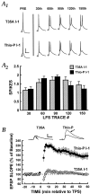
The direct inhibition of postsynaptic PP1 enables TPS-induced LTP without an increase in postsynaptic spiking.A, Postsynaptic activity during TPS was not increased by thiophosphorylated I-1.A1, Samples of postsynaptic potentials recorded just before TPS (PRE) and at every 30th stimulus during TPS are shown. Double spikes were evoked during TPS in both groups. Of all TPS samples recorded (n = 85), only one stimulus produced three spikes, with all of the others yielding one or two. Calibration: 10 mV; 10 msec.A2, Summary of the number of spikes produced by stimuli during TPS is shown. There were no statistically significant differences between cells recorded with thiophosphorylated I-1 and those recorded with T35A I-1. A patterning effect was obtained, with the number of spikes at trace90 significantly higher than those at traces 30 and 150 (Newman–Keuls, p < 0.01 and 0.05, respectively). B, TPS induces LTP when paired with intracellular application of thiophosphorylated I-1. Only cells impaled with Thio-P I-1 (n = 10) showed LTP after TPS. The rapid induction of LTP compared with that shown in Figure 3 is probably caused by the stronger stimulation used during TPS (see Materials and Methods). No lasting effect on synaptic efficiency was seen when TPS was delivered to cells impaled with electrodes containing inactive T35A I-1 (n = 7). The inset shows sample EPSPs obtained during the baseline period and at 60 min after TPS.

Similar articles

Cited by

References

    1. Aston-Jones G, Rajkowski J, Kubiak P, Alexinsky T. Locus coeruleus neurons in monkey are selectively activated by attended cues in a vigilance task. J Neurosci. 1994;14:4467–4480. - PMC - PubMed
    1. Bliss TV, Collingridge GL. A synaptic model of memory: long-term potentiation in the hippocampus. Nature. 1993;361:31–39. - PubMed
    1. Blitzer RD, Wong T, Nouranifar R, Iyengar R, Landau EM. Postsynaptic cAMP pathway gates early LTP in hippocampal CA1 region. Neuron. 1995;15:1403–1414. - PubMed
    1. Blitzer RD, Connor JH, Brown GP, Wong T, Shenolikar S, Iyengar R, Landau EM. Gating of CaMKII by cAMP-regulated protein phosphatase activity during LTP. Science. 1998;280:1940–1942. - PubMed
    1. Braun AP, Schulman H. A non-selective cation current activated via the multifunctional Ca2+-calmodulin-dependent protein kinase in human epithelial cells. J Physiol (Lond) 1995;488:37–55. - PMC - PubMed

Publication types

MeSH terms

Substances

LinkOut - more resources