. 2000 Nov 1;20(21):7932-40.
doi: 10.1523/JNEUROSCI.20-21-07932.2000.

Regulation of AMPA receptor GluR1 subunit surface expression by a 4. 1N-linked actin cytoskeletal association

Affiliations

Regulation of AMPA receptor GluR1 subunit surface expression by a 4. 1N-linked actin cytoskeletal association

L Shen et al. J Neurosci. .

Abstract

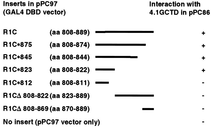
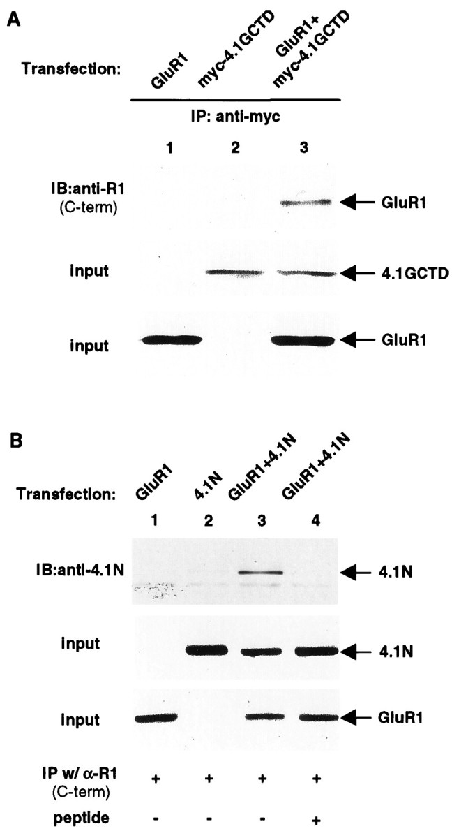
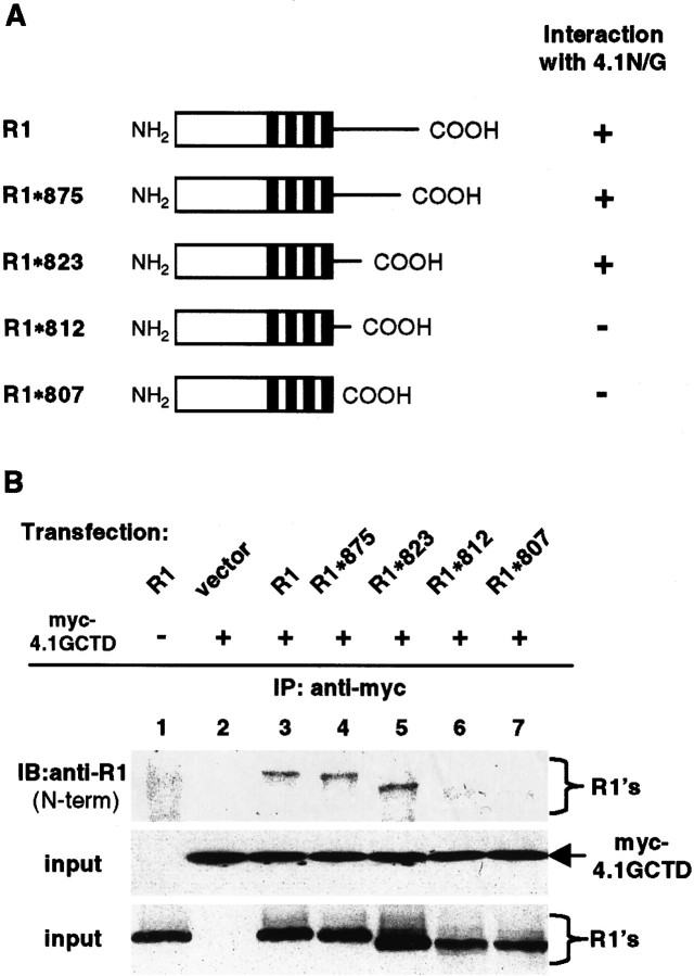
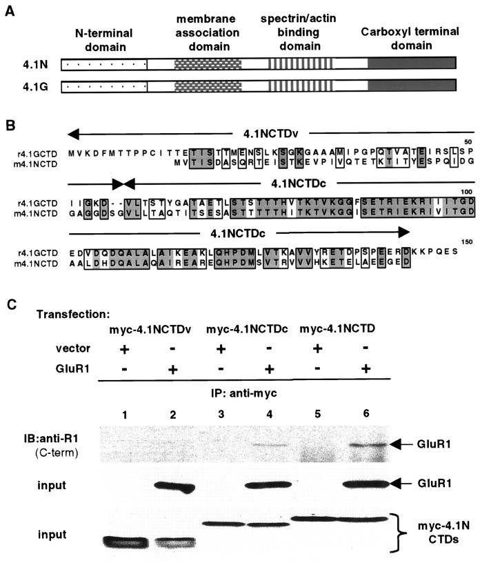
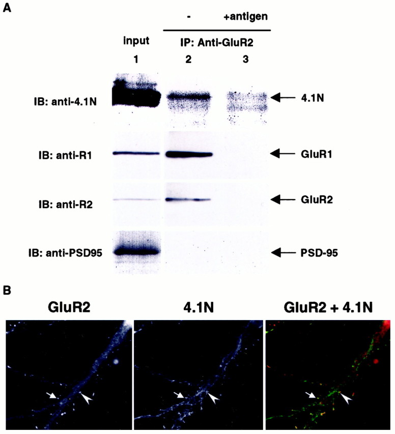
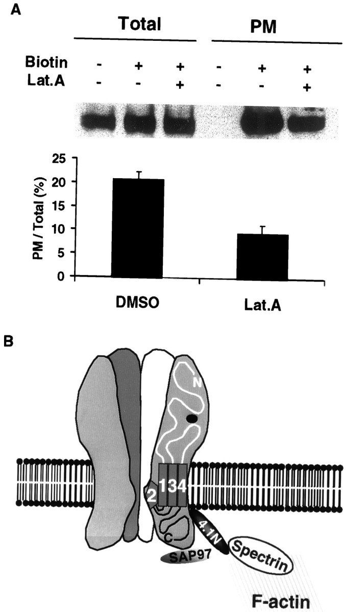
The synaptic localization, clustering, and immobilization of neurotransmitter receptors and ion channels play important roles in synapse formation and synaptic transmission. Although several proteins have been identified that interact with AMPA receptors and that may regulate their synaptic targeting, little is known about the interaction of AMPA receptors with the cytoskeleton. In studies examining the interaction of the AMPA receptor GluR1 subunit with neuronal proteins, we determined that GluR1 interacts with the 4.1G and 4.1N proteins, homologs of the erythrocyte membrane cytoskeletal protein 4.1. Using the yeast two-hybrid system and a heterologous cell system, we demonstrated that both 4.1G and 4.1N bind to a membrane proximal region of the GluR1 C terminus, and that a region within the C-terminal domain of 4.1G or 4.1N is sufficient to mediate the interaction. We also found that 4.1N can associate with GluR1 in vivo and colocalizes with AMPA receptors at excitatory synapses. Disruption of the interaction of GluR1 with 4.1N or disruption of actin filaments decreased the surface expression of GluR1 in heterologous cells. Moreover, disruption of actin filaments in cultured cortical neurons dramatically reduced the level of surface AMPA receptors. These results suggest that protein 4.1N may link AMPA receptors to the actin cytoskeleton.


Figures

Fig. 1.
Fig. 1.
Yeast two-hybrid analysis of GluR1 and protein 4.1G interaction. The cytosolic tail of GluR1 and its various deletions were subcloned into yeast vector pPC97 containing the GAL4 DNA binding domain. The CTD of protein 4.1G was subcloned into pPC86 containing the GAL4 activation domain and co-transformed with GluR1 constructs into yeast. Double transformants were selected and scored for growth on plates lacking leucine, tryptophan, adenine, and histidine and for lacZ activity. Those that tested positive for interaction are designated +; those that tested negative are designated −.
Fig. 2.
Fig. 2.
Association of GluR1 with 4.1G and 4.1N in HEK 293T cells. A. Coimmunoprecipitation of 4.1GCTD with GluR1 from 293T cells. Full-length GluR1 and myc-tagged 4.1GCTD were transfected into 293T cells either individually (lanes 1, 2) or together (lane 3). The transfected cells were solubilized with 1% Triton X-100, and the solubilized cell lysates were immunoprecipitated with anti-myc antibody. The immunoprecipitates were resolved on SDS-PAGE followed by immunoblotting with anti-GluR1 C-terminal antibody (top panel). The presence of myc-tagged 4.1GCTD (middle panel) and GluR1 (bottom panel) in the input for immunoprecipitation was confirmed by immunoblotting with anti-myc and anti-GluR1 C-terminal antibodies, respectively. IP, Immunoprecipitation; IB, immunoblot; same for other legends. B, Coimmunoprecipitation of 4.1N with GluR1 from 293T cells. Full-length GluR1 and full-length 4.1N were transfected into 293T cells either individually (lanes 1, 2) or combined (lanes 3, 4). The immunoprecipitation from cell lysates is the same as inA, except here the anti-GluR1 C-terminal antibody is used. The immunoprecipitates were resolved on SDS-PAGE and probed with anti-4.1N antibody (top panel). As a control experiment in lane 4, the anti-GluR1 C-terminal antibody was preincubated with its specific antigen before immunoprecipitation (see Materials and Methods). The presence of 4.1N (middle panel) and GluR1 (bottom panel) in the input for immunoprecipitation was confirmed by Western blot using anti-4.1N and anti-GluR1 antibodies, respectively.
Fig. 3.
Fig. 3.
Requirement of a membrane proximal region for association of GluR1 with proteins 4.1G and 4.1N. A, Schematic diagram of GluR1 deletion constructs used inB. B, The membrane proximal region in the GluR1 C terminus is required for binding to protein 4.1G. Full-length and C-terminal deletions of GluR1 were transfected with 4.1GCTD, and then solubilized cell lysates were immunoprecipitated with anti-myc antibody. The immunoprecipitates were subjected to SDS-PAGE and Western blot with anti-GluR1 N-terminal antibody (top panel). The presence of myc-tagged 4.1GCTD (middle panel) and GluR1 and its deletions (bottom panel) in the input for immunoprecipitation was confirmed by Western blot using anti-myc and anti-GluR1 N-terminal antibodies, respectively.
Fig. 4.
Fig. 4.
A consensus region within 4.1N is sufficient for interaction with GluR1. A, Schematic diagram of domain structures of 4.1N and 4.1G. The two proteins have significant homology at the membrane association domain, spectrin–actin binding domain, and CTD but have little homology at the N-terminal domain.B, Sequence alignment of CTDs of 4.1N and 4.1G. Identical amino acid residues are boxed anddarkly shaded; conserved residues areboxed and lightly shaded. The alignment shows that CTDs of the two proteins contain a variable N-terminal region (4.1NCTDv) and a highly conserved C-terminal region (4.1NCTDc). C, Association of the consensus region of 4.1NCTD with GluR1. A myc-tagged variable region (myc-4.1NCTDv; lanes 1, 2) or consensus region (myc-4.1NCTDc; lanes 3, 4) of 4.1NCTD or the entire 4.1NCTD (myc-4.1NCTD;lanes 5, 6) was co-transfected with vector (lanes 1, 3, 5) or with GluR1 (lanes 2, 4, 6) into 293T cells. Immunoprecipitations were performed on solubilized cell lysates using anti-myc antibody followed by SDS-PAGE and immunoblotting with anti-GluR1 antibody (top panel). The presence of GluR1 (middle panel) and myc-tagged proteins (bottom panel) in the input for immunoprecipitation was confirmed by Western blot using anti-GluR1 C-terminal and anti-myc antibodies, respectively.
Fig. 5.
Fig. 5.
Protein 4.1N interacts with GluR1 in vivo and colocalizes with AMPA receptor complex in excitatory synapses. A, Rat brain membrane preparation (P2) was solubilized with 1% deoxycholate and immunoprecipitated with either anti-GluR2 N-terminal antibody (lane 2) or the same antibody with antigen preabsorption (lane 3). The immunoprecipitated complex and the input (lane 1) were resolved by SDS-PAGE and probed with anti-4.1N (top panel), anti-GluR1 C-terminal (upper middle panel), anti-GluR2 C-terminal (lower middle panel), and PSD-95 (bottom panel) antibodies. B, Primary hippocampal neurons were double-stained with monoclonal anti-GluR2 antibody (Chemicon, Temecula, CA) and polyclonal anti-4.1N antibody as primary antibodies, followed by rhodamine-conjugated anti-mouse IgG and FITC-conjugated anti-rabbit IgG as secondary antibodies. The staining was visualized and digitized using a fluorescent microscope (Zeiss, Thornwood, NY) with a digital camera controlled by the Metamorph program (Universal Imaging, West Chester, PA). Arrows and arrowheadsindicate the area of colocalization.
Fig. 6.
Fig. 6.
Deletion of the GluR1 4.1 binding region reduces surface expression of GluR1. The membrane proximal region in the GluR1 C terminus is important for receptor surface expression. COS cells were transfected with pRK5 (Mock), GluR1*812 (R1*812), or GluR1*823 (R1*823). Surface expression of GluR1 was determined by biotinylation, as described in Materials and Methods. Samples from total cell lysates (Total) and biotinylated samples (PM) were analyzed for GluR1. For quantitation, the signals from biotinylated samples were divided by the total GluR1 signals to obtain the ratio of surface expression for GluR1*823 (R1*823) and GluR1*812 (R1*812). The ratio of surface expression of GluR1*812 was normalized to that of GluR1*823 to obtain the relative value of surface expression. n = 3. The error bar indicates SEM.
Fig. 7.
Fig. 7.
F-actin regulates GluR1 surface expression in COS cells. A, Disruption of F-actin inhibits surface expression of GluR1 in COS cells. COS cells were transiently transected with WT GluR1. Twenty-four hours after the transfection, either DMSO or a concentrated DMSO stock of latrunculin A (Lat.A) was added to the culture medium. Final concentration of latrunculin A is 5 μm. Cells were further incubated at 37°C for 2 hr before cell surface biotinylation. Samples from cell lysates (Total) and biotinylated samples (PM) were analyzed for GluR1 using an anti-GluR1 N-terminal antibody. For quantitation, the biotinylated samples were normalized to total GluR1 signals to obtain the ratio of surface expression for the control and latrunculin-treated cells. The ratio of surface expression of the latrunculin-treated cells was normalized to that of control cells to express the relative surface expression. n = 3. The error bar indicates SEM.B. The C-terminal membrane proximal region of GluR1 is important for latrunculin inhibition of surface expression. COS cells transfected with pRK5 (Mock), R1*812, or R1*823 were treated with either DMSO (Lat.A −) or latrunculin A (Lat.A +), as described in Materials and Methods. For quantitation, the signals from biotinylated samples were normalized to the total GluR1 signal to obtain the ratio of surface expression. The ratio of the surface expression was then normalized to that of R1*823 without latrunculin. R1*823,R1*823+Lat.A, R1*823 without or with latrunculin A, respectively;R1*812,R1*812+Lat.A, R1*812 without or with latrunculin A, respectively. n = 3. Error bars indicate SEM.
Fig. 8.
Fig. 8.
Overexpression of CTDs of 4.1 attenuated GluR1 surface expression. COS cells were cotransfected using R1*812 or R1*823, together with pRK5 (vector), 4.1GCTD (myc-4.1GCTD), or 4.1NCTD (myc-4.1NCTD). Both CTDs were myc-tagged. Transfected cells were incubated at 37°C for 24 hr, followed by cell surface biotinylation, as described in Materials and Methods. A, Immunoblots. Top panel, Total cell lysates blotted with anti-myc antibody;middle panel, total cell lysates blotted with anti-GluR1 N-terminal antibody; bottom panel, biotinylated samples blotted with anti-GluR1 N-terminal antibody. B, Quantitation of surface expression. The signals from biotinylated samples were normalized to the total GluR1 signal to obtain the ratio of surface expression and then normalized to the surface expression of GluR1*823 to obtain the relative value of surface expression.n = 3. Error bars indicate SEM.
Fig. 9.
Fig. 9.
Actin cytoskeleton is required for synaptic maintenance of AMPA receptors. A, Actin polymerization is required for synaptic maintenance of AMPA receptors. Cultured rat hippocampal neurons were treated with either DMSO (Lat.A−) or latrunculin A (Lat.A +) followed by cell surface biotinylation (Biotin +, −) as described in Materials and Methods. Samples from cell lysates (Total) and biotinylated samples (PM) were analyzed for GluR1 via anti-GluR1 antibody. Samples of immunoblots (top panel) and a summary of quantitation (bottom panel) are presented. For quantitation, the signals from biotinylated samples were normalized to total GluR1 signals to obtain the ratio of surface expression [PM/Total (%)].DMSO, Lat.A, GluR1 without or with latrunculin A, respectively. n = 4. Error bars indicate SEM.B, Schematic model of AMPA receptor–actin cytoskeleton cross-linking by 4.1N. The interaction of the GluR1 subunit with protein 4.1N and SAP97 may link surface AMPA receptors to the cortical actin cytoskeleton network underneath the synaptic plasma membrane and PSD.

References

    1. Allison DW, Gelfand VI, Spector I, Craig AM. Role of actin in anchoring postsynaptic receptors in cultured hippocampal neurons: differential attachment of NMDA versus AMPA receptors. J Neurosci. 1998;18:2423–2436. - PMC - PubMed
    1. Anderson RA, Lovrien RE. Glycophorin is linked by band 4.1 protein to the human erythrocyte membrane skeleton. Nature. 1984;307:655–658. - PubMed
    1. Bennett V, Gilligan DM. The spectrin-based membrane skeleton and micron-scale organization of the plasma membrane. Annu Rev Cell Biol. 1993;9:27–66. - PubMed
    1. Blackstone CD, Levey AI, Martin LJ, Price DL, Huganir RL. Immunological detection of glutamate receptor subtypes in human central nervous system. Ann Neurol. 1992a;31:680–683. - PubMed
    1. Blackstone CD, Moss SJ, Martin LJ, Levey AI, Price DL, Huganir RL. Biochemical characterization and localization of a non-N-methyl-D-aspartate glutamate receptor in rat brain. J Neurochem. 1992b;58:1118–1126. - PubMed

Publication types

MeSH terms

LinkOut - more resources