. 2000 Nov 1;20(21):8005-11.
doi: 10.1523/JNEUROSCI.20-21-08005.2000.

NMDA but not non-NMDA excitotoxicity is mediated by Poly(ADP-ribose) polymerase

Affiliations

NMDA but not non-NMDA excitotoxicity is mediated by Poly(ADP-ribose) polymerase

A S Mandir et al. J Neurosci. .

Abstract

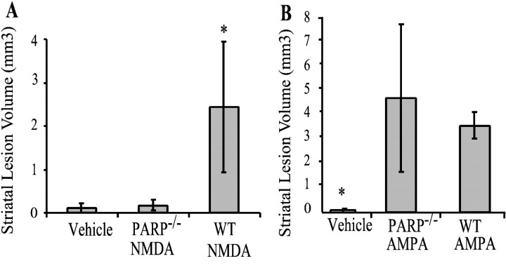
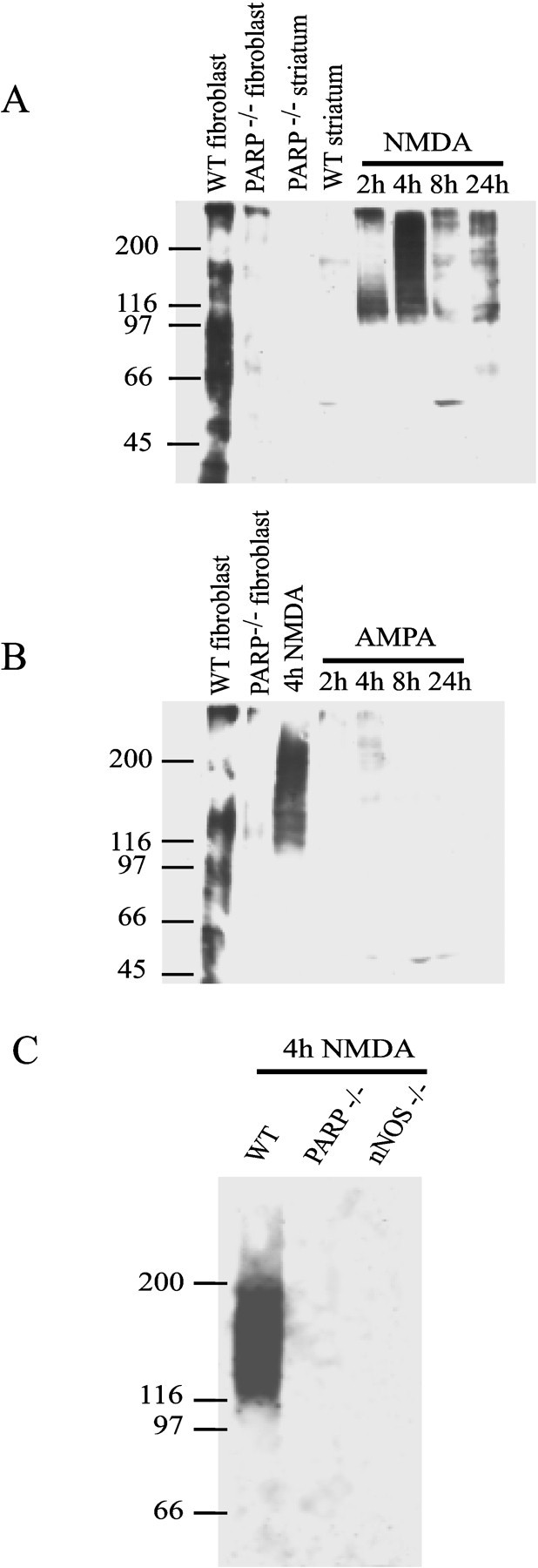
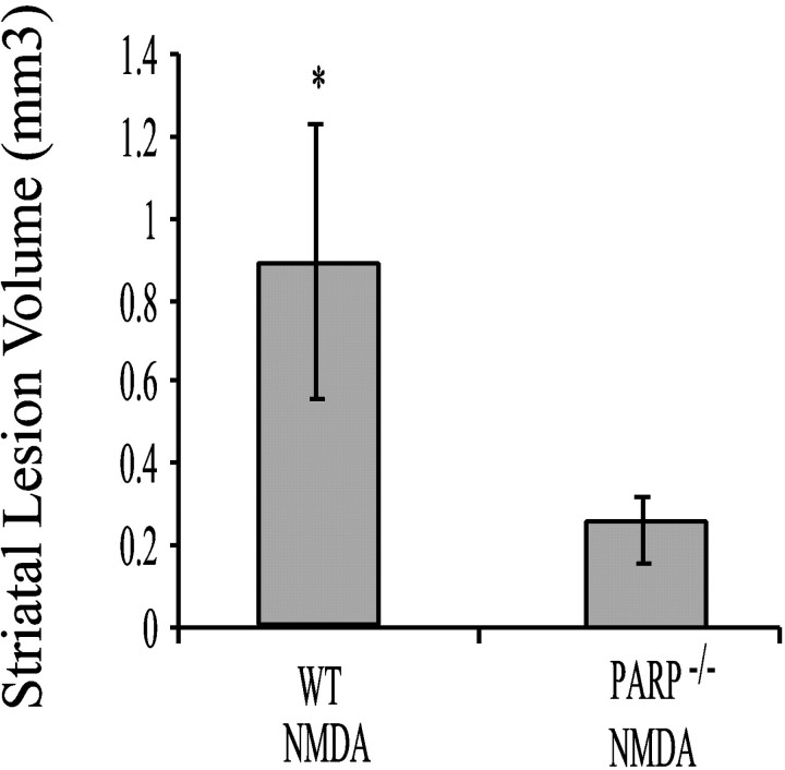
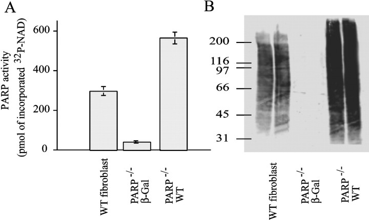
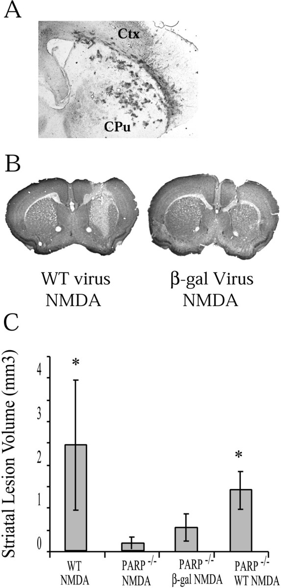
Poly(ADP-ribose) polymerase (PARP-1), a nuclear enzyme that facilitates DNA repair, may be instrumental in acute neuronal cell death in a variety of insults including, cerebral ischemia, 1-methyl-4-phenyl-1,2,3,6-tetrahydropyridine-induced parkinsonism, and CNS trauma. Excitotoxicity is thought to underlie these and other toxic models of neuronal death. Different glutamate agonists may trigger different downstream pathways toward neurotoxicity. We examine the role of PARP-1 in NMDA- and non-NMDA-mediated excitotoxicity. NMDA and non-NMDA agonists were stereotactically delivered into the striatum of mice lacking PARP-1 and control mice in acute (48 hr) and chronic (3 week) toxicity paradigms. Mice lacking PARP-1 are highly resistant to the excitoxicity induced by NMDA but are as equally susceptible to AMPA excitotoxicity as wild-type mice. Restoring PARP-1 protein in mice lacking PARP-1 by viral transfection restored susceptibility to NMDA, supporting the requirement of PARP-1 in NMDA neurotoxicity. Furthermore, Western blot analyses demonstrate that PARP-1 is activated after NMDA delivery but not after AMPA administration. Consistent with the theory that nitric oxide (NO) and peroxynitrite are prominent in NMDA-induced neurotoxicity, PARP-1 was not activated in mice lacking the gene for neuronal NO synthase after NMDA administration. These results suggest a selective role of PARP-1 in glutamate excitoxicity, and strategies of inhibiting PARP-1 in NMDA-mediated neurotoxicity may offer substantial acute and chronic neuroprotection.


Figures

Fig. 1.
Fig. 1.
Representative coronal sections stained with cresyl violet to reveal lesions in WT (A,C, E) and PARP-1−/−(B, D, F) mice 48 hr after treatment with NMDA, AMPA, or vehicle alone. PARP-1−/− mice demonstrate remarkable resistance to NMDA intrastriatal injections with lesion volumes similar to vehicle alone. Lesion volumes in PARP-1−/− are not significantly different from WT mice receiving intrastriatal injections of AMPA.
Fig. 2.
Fig. 2.
Average lesions volumes from WT and PARP-1−/− mice 48 hr after 20 nmol of NMDA (A) (n = 7) or 6 nmol of AMPA (B) (n = 6). Volumes are expressed as mean ± SD. In A,asterisk indicates significant difference between PARP-1−/− and WT after NMDA injections (p < 0.5; ANOVA). No statistical differences are observed between PARP-1−/− and WT animals receiving intrastriatal AMPA injections. In B,asterisk indicates both are significantly different than vehicle injection lesion volumes.
Fig. 3.
Fig. 3.
Cortical cultures from WT or PARP-1−/−(P−/−) express equal levels of NMDA-R, GluR1, and GluR2/3 as determined by Western blot analysis. The experiment was performed twice with similar results. Cortical cultures from WT or PARP-1−/− mice exposed to NMDA (500 μm), vehicle alone, and AMPA (50 μm) with or without the presence of the glutamate antagonist DNQX. PARP-1−/− or WT cultures exposed to AMPA alone demonstrate significant percentage of cell death compared with control or DNQX-treated cultures (p < 0.01; ANOVA; mean ± SD). Unlike the substantial protection exhibited by PARP-1−/− cultures to NMDA neurotoxicity compared with WT as shown previously (Eliasson et al., 1997), PARP-1−/− cultures demonstrate a partial but significant percentage of protection to AMPA neurotoxicityin vitro (*p < 0.05, WT compared with PARP knock-outs; significance determined by ANOVA with Newman–Keuls post hoc analysis). Experiments were replicated a minimum of two times with at least 8000–20,000 neurons counted per experiment.
Fig. 4.
Fig. 4.
A, Western blots demonstrating the time course of PARP-1 activation after 20 nmol of NMDA intrastriatal injections in WT mice. Striata from PARP-1−/− or WT mice were prepared as described in Materials and Methods. PAR polymer formation is seen 2 hr after injection with a peak detection of polymer at 4 hr after injection. Sonicated fibroblasts incubated with NAD serve as the positive control. B, Time course of PARP-1 activation after 6 nmol of AMPA intrastriatal injection in wild-type mice. Minimal PARP-1 activation is seen compared with NMDA injections (4 hr NMDA PAR formation shown for comparison).C, Although PARP-1 is activated maximally at 4 hr after NMDA injection in WT mice, nNOS−/− mice do not demonstrate PAR polymer formation after NMDA injection. These results were replicated three times with similar results.
Fig. 5.
Fig. 5.
Histogram of mean intrastriatal volumes 3 weeks after NMDA injection in WT (n = 3) and PARP-1−/− (n = 3) mice. PARP-1−/− mice still demonstrate minimal lesion volume chronically after NMDA injection. WT mice demonstrate significantly greater lesion volumes than PARP-1−/− mice (*p < 0.05; Student's t test).
Fig. 6.
Fig. 6.
PARP-1 activity in WT and PARP-1−/− fibroblast cells infected with Sindbis virus-containing cDNA encoding lacZ or WT PARP-1.A, WT or PARP-1−/− fibroblasts infected with WT PARP-1 demonstrate prominent PARP-1 activation as quantified by amount of [32P]NAD incorporated after sonication in the presence of [32P]NAD.B, PAR polymer formation determined by Western blot indicates that wild-type PARP-1 Sindbis virus restores PARP-1 activity in PARP-1−/− fibroblasts. Data shown are representative of duplicate experiments.
Fig. 7.
Fig. 7.
A, Coronal section through striatum 4 d after intrastriatal injection of Sindbis virus-expressinglacZ. Virus is prominent in part of the striatum. These results were replicated at least three times. B, Coronal section of a PARP-1−/− mouse receiving Sindbis virus-expressing cDNA of WT PARP-1 and then 48 hr after an intrastriatal injection of 20 nmol of NMDA. The section is stained with cresyl violet and demonstrates a demarcated area of NMDA damage.C, Lesion volumes (mean ± SD) of PARP-1−/− receiving Sindbis virus encoding PARP-1 WT cDNA (n = 3) demonstrate lesion volumes approaching that of WT mice receiving NMDA. PARP-1−/− mice receiving Sindbis virus only encoding lacZ (n = 3) demonstrate significantly lower lesion volumes (significance determined by ANOVA with Newman–Keuls post hoc analysis; *p < 0.05, significantly different from PARP-1−/− NMDA).

References

    1. Affar EB, Duriez PJ, Shah RG, Sallmann FR, Bourassa S, Kupper JH, Burkle A, Poirier GG. Immunodot blot method for the detection of poly (ADP-ribose) synthesized in vitro and in vivo. Anal Biochem. 1998;259:280–283. - PubMed
    1. Affar EB, Duriez PJ, Shah RG, Winstall E, Germain M, Boucher C, Bourassa S, Kirkland JB, Poirier GG. Immunological determination and size characterization of poly(ADP- ribose) synthesized in vitro and in vivo. Biochim Biophys Acta. 1999;1428:137–146. - PubMed
    1. Ayata C, Ayata G, Hara H, Matthews RT, Beal MF, Ferrante RJ, Endres M, Kim A, Christie RH, Waeber C, Huang PL, Hyman BT, Moskowitz MA. Mechanisms of reduced striatal NMDA excitotoxicity in type I nitric oxide synthase knock-out mice. J Neurosci. 1997;17:6908–6917. - PMC - PubMed
    1. Beckman JS, Crow JP. Pathological implications of nitric oxide, superoxide and peroxynitrite formation. Biochem Soc Trans. 1993;21:330–334. - PubMed
    1. Berger NA. Poly(ADP-ribose) in the cellular response to DNA damage. Radiat Res. 1985;101:4–15. - PubMed

Publication types

MeSH terms

Substances

LinkOut - more resources