. 2000 Nov 1;20(21):8061-8.
doi: 10.1523/JNEUROSCI.20-21-08061.2000.

Regeneration of lesioned corticospinal tract fibers in the adult rat induced by a recombinant, humanized IN-1 antibody fragment

Affiliations

Regeneration of lesioned corticospinal tract fibers in the adult rat induced by a recombinant, humanized IN-1 antibody fragment

C Brösamle et al. J Neurosci. .

Abstract

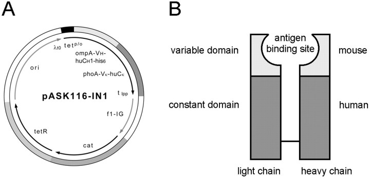
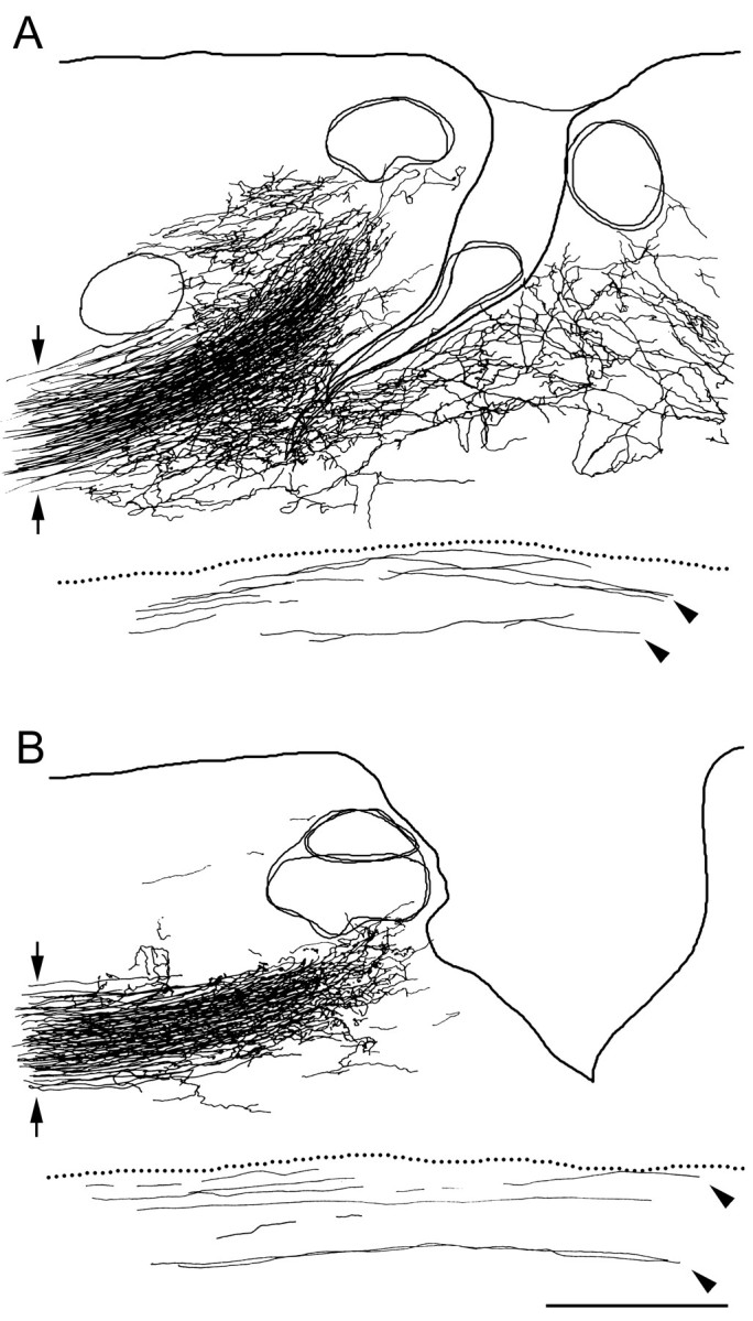
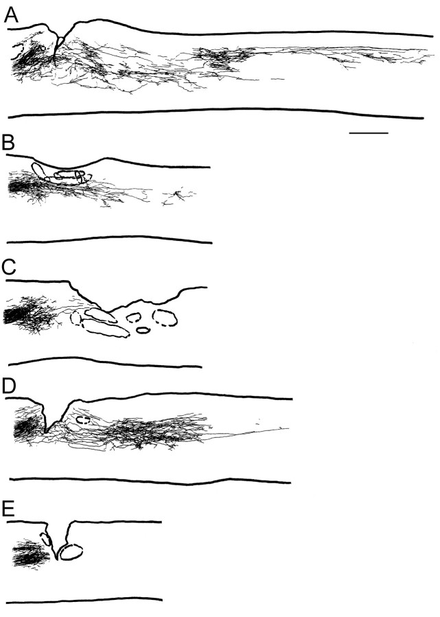
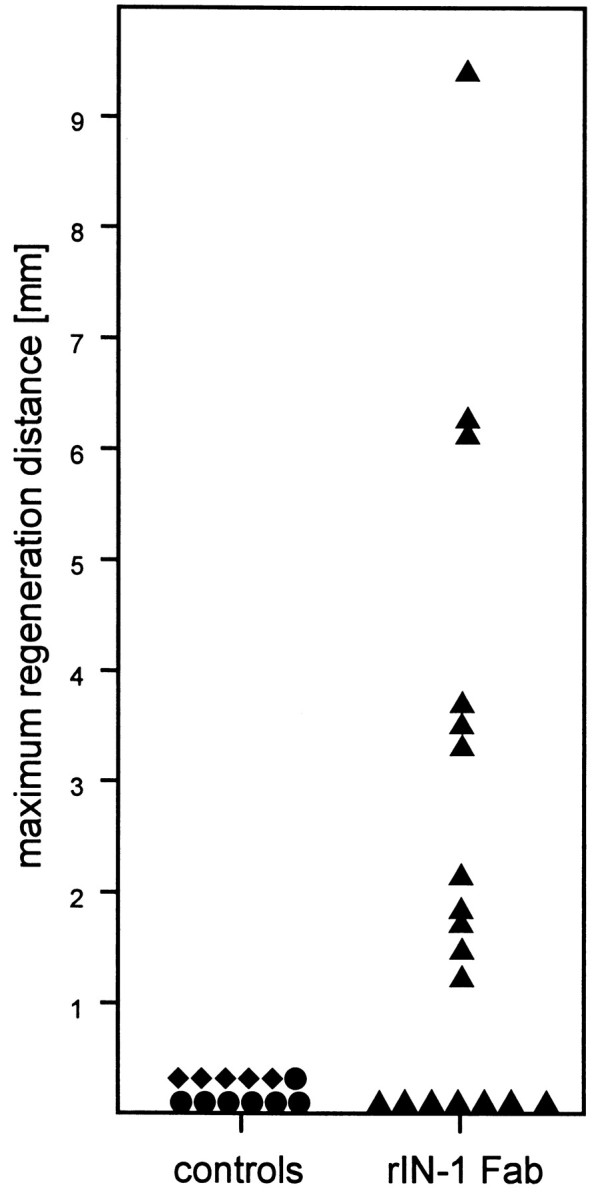
Axons in the CNS of higher vertebrates generally fail to regenerate after injury. This lack of regeneration is crucially influenced by neurite growth inhibitory protein constituents of CNS myelin. We have shown previously that a monoclonal antibody (mAb IN-1) capable of binding and neutralizing Nogo-A, a myelin-associated inhibitor of neurite growth, can induce long-distance axonal regeneration and increased structural plasticity with improved functional recovery in rat models of CNS injury. In this paper we demonstrate that a partially humanized, recombinant Fab fragment (rIN-1 Fab) derived from the original mAb IN-1, was able to promote long-distance regeneration of injured axons in the spinal cord of adult rats. When infused into a spinal cord injury site, regrowth of corticospinal fibers in 11 of 18 animals was observed after a survival time of 2 weeks. Regenerating fibers grew for >9 mm beyond the lesion site and arborized profusely in the distal cord. Regenerated fibers formed terminal arbors with varicosities in the spinal cord gray matter, strongly resembling synaptic points of contact to neurons in the spinal cord distal to the lesion. In animals that had received a bovine serum albumin solution or a recombinant IN-1 fragment that had been mutated in the antigen binding site (mutIN-1 Fab), no significant growth beyond normal lesion-induced sprouting was observed. Neutralization of endogenous nerve growth inhibitors represents a novel use of recombinant antibody technology with potential therapeutic applications after traumatic CNS lesions.


Figures

Fig. 1.
Fig. 1.
Recombinant, humanized IN-1 Fab. A, Expression vector pASK116-IN1. The two chains of the Fab fragment are fused with the bacterial OmpA and PhoA signal sequences, respectively, and the heavy chain carries a His6 tag at its C terminus. The structural genes for both polypeptide chains are arranged in a dicistronic operon under transcriptional control of thetet promoter/operator (tetp/o), ending with the lipoprotein terminator (tlpp). Tight repression of the tet promoter is controlled by the tetrepressor gene (tetR). ori, Origin of replication; λt0, phage λ transcription terminator; f1-IG, intergenic region of filamentous phage f1; cat, Cm resistance gene.B, The recombinant IN-1 Fab carries the variable domains of the original (murine) IgM/κ mAb IN-1 determining the antigen specificity and constant domains of human origin, IgG1/κ subclass.
Fig. 2.
Fig. 2.
rIN-1 Fab stains CNS white matter and recognizes recombinant Nogo-A. A, Recombinant, humanized IN-1 Fab stains myelinated structures in the CNS. Unmyelinated areas such as the superficial laminae of the spinal cord and peripheral myelinated tissues (dorsal and ventral roots, asterisks) are not or only weakly stained. This staining pattern very closely resembles that of the original IN-1 mAb. B, Sections incubated with the secondary antibody only were not significantly stained. Scale bar, 300 μm. C, Recombinant, full-length Nogo-A prepared from stably transfected CHO cells is recognized by rIN-1 Fab on Western blot as shown by reprobing the same blot with an anti-myc antibody to reveal recombinant protein.
Fig. 3.
Fig. 3.
Humanized rIN-1 Fab recognizes and neutralizes myelin-associated neurite growth inhibitors in vitro.A, 3T3 fibroblast spreading is inhibited when the cells are plated on a CNS myelin protein substrate enriched for Nogo-A (q-pool) (Spillmann et al., 1997, 1998). When the inhibitory substrate is pretreated with mAb IN-1 hybridoma supernatant (50 μg/ml IgM), inhibition is reduced drastically. rIN-1 Fab has a similar neutralizing effect at concentrations of 500–50 μg/ml and is less efficient at higher dilutions. Mutated rIN-1 Fab did not show neutralizing activity. **p < 0.01, Student'st test, compared with q-pool (100%). B, Neurite outgrowth from E16 chick dorsal root ganglia is strongly inhibited when the DRGs are plated on q-pool. Pretreatment with humanized rIN-1 Fab allows for outgrowth of large numbers of long neurites within 24 hr. Mutated rIN-1 Fab did not neutralize Nogo-A in the q-pool substrate. Neurite outgrowth was scored in arbitrary units from 0 (no outgrowth) to 4 (maximal outgrowth on a laminin substrate). *p < 0.05, Student's t test, compared with q-pool. Scale bar, 30 μm. Error bars indicate means ± SEM.
Fig. 4.
Fig. 4.
Spinal cord injury model and subdural antibody infusion. A, Laminectomies were performed at levels T8 and T10. With fine iridectomy scissors, the dorsal half of the spinal cord was cut bilaterally at T8, thus transecting the main dorsomedial and the minor dorsolateral corticospinal tract components. Through a small hole in the dura a catheter connected to an osmotic mini-pump was inserted into the subdural space close to the lesion at T8 to infuse the lesion area and the distal cord with rIN-1 Fab solution.B, Distribution of infused rIN-1 Fab was controlled by labeling the antibodies with the fluorochrome Alexa488. After 1 week of infusion, rIN-1 Fab had spread well at the infusion site, around the lesion (asterisk), and in the distal cord. rIN-1 Fab concentration was higher in gray matter, but Alexa488 fluorescence could also be easily detected in white matter. Macrophages that had infiltrated the lesion (arrowheads) exhibited strong blue-green fluorescence caused by uptake of the infused rIN-1 Fab and yellowish-green autofluorescence. Scale bar, 500 μm.
Fig. 5.
Fig. 5.
Two examples of BDA-labeled corticospinal fibers at the lesion site in animals treated with rIN-1. Sagittal 50 μm vibratome sections. A, Overview of a typical lesion area in a rIN-1 Fab-treated animal 2 weeks after injury. The labeled corticospinal tract, which runs at the medioventral aspect of the dorsal funiculus in the rat, approaches the lesion from rostral (left) and is interrupted at the lesion. Cellular infiltrates and cavities are present (asterisks).Arrows indicate regenerated fibers that have circumvented the lesion area and are growing in a typical curved and irregular pattern away from the lesion in the caudal direction.B, A close-up in a different rIN-1 animal shows that regenerating fibers growing around and through the lesion are very tortuous and curved. Scale bar (shown in B):A, 100 μm; B, 25 μm.
Fig. 6.
Fig. 6.
Camera lucida reconstructions of consecutive sections fibers at the lesion site in an animal infused with rIN-1 Fab (A) and a control animal infused with mut rIN-1 Fab (B). In the rIN-1 Fab-infused animal, strong sprouting of the lesioned CST fibers can be observed. Some of these sprouts elongate and grow around and through the lesion and farther down the cord. In the mut rIN-1-infused animal, some sprouts have emanated from the lesioned CST, but no long-distance growth occurs. The number of labeled CST fibers and their labeling intensity (arrows), lesion depth, and size are similar in the two animals. Scale bar, 500 μm.
Fig. 7.
Fig. 7.
Regenerated corticospinal fibers in the spinal cord distal to the lesion site branch profusely and give rise to large arborizations in intermediate laminae of the cord (A,B). Higher magnification (C–F) reveals that these arborizations are decorated with many varicosities (arrowheads) that strongly resemble presynaptic boutons in normal corticospinal innervation of spinal gray matter (G). Both terminal and en passant presynaptic boutons can be identified. Parasagittal sections are 50 μm thick. Scale bar (shown in G): A,B, 250 μm; C, D, 30 μm; E–G, 15 μm.
Fig. 8.
Fig. 8.
Camera lucida reconstructions of consecutive series of parasagittal sections (50 μm thick) of the thoracic and high lumbar spinal cord of four representative rIN-1 Fab-treated animals (A–D) and a representative control animal (E). Most rIN-1 Fab-treated animals exhibited long-distance regeneration for up to >9 mm with axons branching profusely into gray matter areas, whereas control animals showed no regenerative growth. Scale bar, 1 mm.
Fig. 9.
Fig. 9.
Maximal elongation of regenerating axons in response to control (●, BSA; ♦, mut rIN-1 Fab) or rIN-1 Fab (▴) treatment. Every symbol represents one animal and indicates the distance of the longest regenerated CST fiber from the center of the lesion. Although in the rIN-1 Fab group 11 of 18 animals showed considerable regrowth of transected CST fibers, this was never observed in control animals.

References

    1. Bandtlow C, Schiweck W, Tai HH, Schwab ME, Skerra A. The Escherichia coli-derived Fab fragment of the IgM/kappa antibody IN-1 recognizes and neutralizes myelin-associated inhibitors of neurite growth. Eur J Biochem. 1996;241:468–475. - PubMed
    1. Beattie MS, Bresnahan JC, Komon J, Tovar CA, Van Meter M, Anderson DK, Faden AI, Hsu CY, Noble LJ, Salzman S, Young W. Endogenous repair after spinal cord contusion injuries in the rat. Exp Neurol. 1997;148:453–463. - PubMed
    1. Bregman BS, Kunkel-Bagden E, Schnell L, Dai HN, Gao D, Schwab ME. Recovery from spinal cord injury mediated by antibodies to neurite growth inhibitors. Nature. 1995;378:498–501. - PubMed
    1. Brösamle C, Schwab ME. Cells of origin, course, and termination patterns of the ventral, uncrossed component of the mature rat corticospinal tract. J Comp Neurol. 1997;386:293–303. - PubMed
    1. Cadelli D, Schwab ME. Regeneration of lesioned septohippocampal acetylcholinesterase-positive axons is improved by antibodies against the myelin-associated neurite growth inhibitors NI-35/250. Eur J Neurosci. 1991;3:825–832. - PubMed

Publication types

MeSH terms