. 2000 Nov 1;20(21):8103-10.
doi: 10.1523/JNEUROSCI.20-21-08103.2000.

Muscarinic tone sustains impulse flow in the septohippocampal GABA but not cholinergic pathway: implications for learning and memory

Affiliations

Muscarinic tone sustains impulse flow in the septohippocampal GABA but not cholinergic pathway: implications for learning and memory

M Alreja et al. J Neurosci. .

Abstract

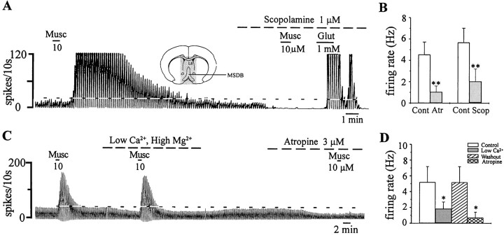
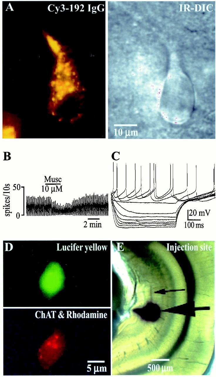
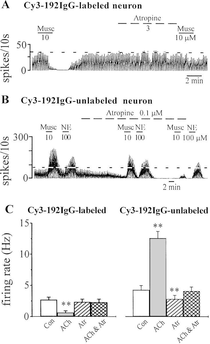
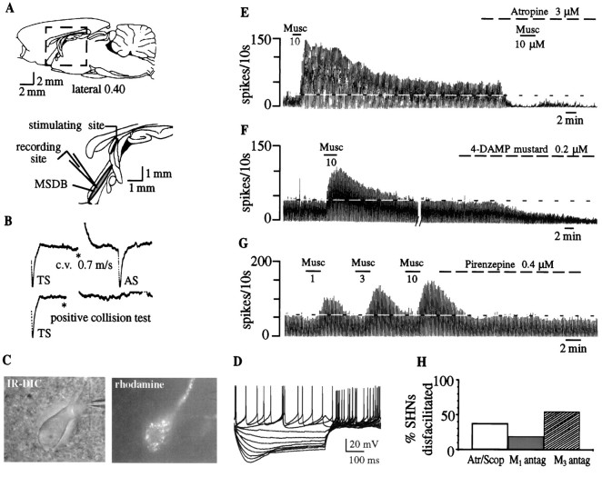
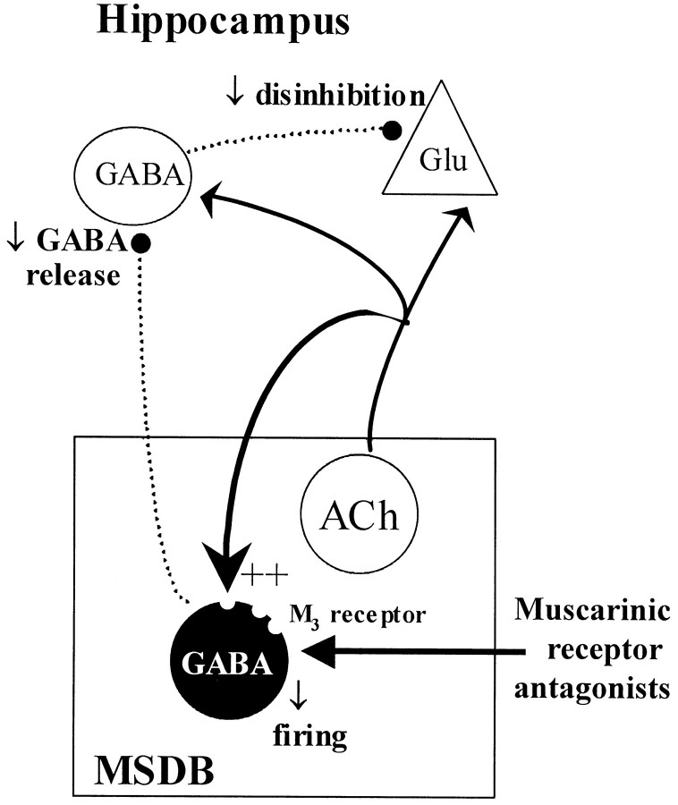
Systemic infusions of the muscarinic cholinergic receptor antagonists atropine and scopolamine (atr/scop) produce an amnesic syndrome in humans, subhuman primates, and rodents. In humans, this syndrome may resemble early symptoms of Alzheimer's disease. Behavioral studies in rats have demonstrated that the medial septum/diagonal band of Broca (MSDB), which sends cholinergic and GABAergic projections to the hippocampus, is a critical locus in mediating the amnesic effects of atr/scop. The amnesic effects of atr/scop in the MSDB have been presumed but not proven to be caused by a decrease in hippocampal acetylcholine (ACh) release after blockade of a muscarinic tone in the MSDB. Using electrophysiological recordings and fluorescent-labeling techniques to identify living septohippocampal neurons in rat brain slices, we now report that, contrary to current belief, a blockade of the muscarinic tone in the MSDB does not decrease impulse flow in the septohippocampal cholinergic pathway; instead, it decreases impulse flow in the septohippocampal GABAergic pathway via M(3) muscarinic receptors. We also report that the muscarinic tone in the MSDB is maintained by ACh that is released locally, presumably via axon collaterals of septohippocampal cholinergic neurons. As such, cognitive deficits that occur in various neurodegenerative disorders that are associated with a loss or atrophy of septohippocampal cholinergic neurons cannot be attributed solely to a decrease in hippocampal acetylcholine release. An additional, possibly more important mechanism may be the concomitant decrease in septohippocampal GABA release and a subsequent disruption in disinhibitory mechanisms in the hippocampus. Restoration of impulse flow in the septohippocampal GABA pathway, possibly via M(3) receptor agonists, may, therefore, be critical for successful treatment of cognitive deficits associated with neurodegenerative disorders such as Alzheimer's and Parkinson's disease.


Figures

Fig. 1.
Fig. 1.
A muscarinic tone is present in the MSDB in vitro. A, Extracellular recording from an MSDB neuron shows that muscarine (Musc) produced a profound increase in firing rate. Scopolamine, a muscarinic receptor antagonist, reduced the basal firing rate and blocked the effect of muscarine; the cell still responded to glutamate (Glut) with a strong excitation. Inset, A coronal section of the brain at the level of the septal nucleus is shown. The slice preparation was restricted to the stippled area. B, A summary is shown of the effect of atropine (Atr) and scopolamine (Scop) on the basal firing rate of neurons that responded to ACh with an increase in firing rate. Thus, MSDB neurons are under a constant muscarinic tone, presumably because of the presence of locally released ACh. C, A blockade of synaptic transmission, using a low-Ca2+, high-Mg2+-containing external solution, also reduced basal firing rates but did not block the excitatory effects of exogenously applied agonist. Atropine both reduced basal firing and blocked the excitatory effects of muscarine. D, A summary of the effect of blocking synaptic transmission on basal firing rates in MSDB neurons is shown. Cont, Control. *p < 0.05. Horizontal dashed lineindicates baseline firing.
Fig. 2.
Fig. 2.
Spontaneously firing cholinergic neurons are present in the MSDB in vitro in brain slices.A, Left, A Cy3–192IgG-labeled neuron visualized in a living brain slice is shown. Cy3–192IgG labels only cholinergic neurons within the MSDB (Hartig et al., 1998; Wu et al., 2000). Right, An IR-DIC image of the same cell shows its healthy appearance. B, Extracellular recordings from a spontaneously firing Cy3–192IgG-labeled neuron that was inhibited by muscarine are shown. Fifty-five percent of the Cy3-labeled cholinergic neurons recorded from fired spontaneously. C, Electrophysiological signature of a Cy3–192IgG-labeled neuron obtained in response to depolarizing and hyperpolarizing pulses (step size, 0.2 nA; maximum step, +0.4 nA) is shown. D, A triple-labeled septohippocampal cholinergic neuron that also fired spontaneously (data not shown) is shown. The cell was labeled with Lucifer yellow after establishment of whole-cell recording. Subsequently, the cell also tested positive for choline acetyltransferase (diffuse red stain), a marker of cholinergic neurons. E, This neuron also colocalized the retrograde marker rhodamine beads (red dots marked by arrows), which were injected into the hippocampus in vivo, 2 d before slicing.
Fig. 3.
Fig. 3.
Effect of muscarinic receptor antagonists on Cy3–192IgG-labeled and unlabeled MSDB neurons. A, Recording from a Cy3–192IgG-labeled cholinergic neuron shows that although atropine (3 μm) had no effect on the baseline firing rate, it did block the inhibitory effect of exogenously applied muscarine. B, In contrast, in a Cy3–192IgG-unlabeled neuron (presumably, GABAergic), a low nanomolar concentration of atropine reduced basal firing rates and also blocked the effect of exogenous muscarine but had little effect on norepinephrine (NE)-induced excitation; NE has been shown previously to excite septohippocampal GABA neurons (Alreja and Liu, 1996). C, The bar chart summarizes the effects of muscarinic drugs on Cy3–192IgG-labeled (i.e., cholinergic) and Cy3–192IgG-unlabeled neurons. Note that atropine had no effect on Cy3-labeled neurons; unlabeled neurons that are excited by muscarine show a decrease in basal firing rates in the presence of atropine.Con, Control. **p < 0.005.
Fig. 4.
Fig. 4.
Effect of muscarinic receptor antagonists on septohippocampal neurons. A, Top, Sagittal section through the rat brain shows the septal area.Bottom, The boxed area in the top panel is enlarged and shows the MSDB that was the recording site. For antidromic identification of septohippocampal neurons, the stimulating electrode was placed in the dorsal fornix because it conveys both cholinergic and GABAergic MSDB fibers to the hippocampus.B, Top, Extracellular recording from a spontaneously firing, antidromically activated septohippocampal neuron is shown. A spontaneous spike was used to trigger the oscilloscope (TS), the dorsal fornix was stimulated (*) 4 msec later, and an antidromically activated spike (AS) was obtained after a latency of 3.3 msec. This cell was classified as a GABA type on the basis of the calculated conduction velocity of 0.7 m/sec.Bottom, A positive collision test, in which the cell could not be activated antidromically when the dorsal fornix was stimulated 3 msec after the triggering spike, is shown. For additional criteria used for confirming antidromicity, see Alreja and Liu (1996).C, An IR-DIC image is shown of a septohippocampal neuron identified using the retrograde tracer rhodamine beads that were injected into the hippocampus 2 d before recording.D, Electrophysiological signature of a rhodamine-labeled neuron is obtained in response to depolarizing and hyperpolarizing pulses (step size, 0.2 nA; maximum step, +0.4 nA). Note the depolarizing sag that is characteristic of septohippocampal GABA but not cholinergic neurons. This neuron was excited by muscarine (data not shown), a property that is exclusive to noncholinergic MSDB neurons.E, The chart record shows that bath-applied muscarine produced a profound and prolonged increase in the firing rate in an SHN. Atropine, a muscarinic receptor antagonist, reduced basal firing and blocked the response to a subsequent application of muscarine.F, Low nanomolar concentrations of 4-DAMP mustard, an M3-selective antagonist, reduced basal firing and also blocked the effects of exogenous muscarine (data not shown).G, The concentration-dependent excitatory effect of muscarine in another SHN is shown. Pirenzepine, an M1-selective antagonist, had no effect on the basal firing rate. H, The effects of antagonists are summarized. Although atr/scop and the M3-selective antagonist reduced basal firing rates in 40–50% of the neurons tested, the M1 antagonist had an effect in 20% of the neurons tested.Antag, Antagonist.
Fig. 5.
Fig. 5.
Schematic figure shows that ACh released via axon collaterals of septohippocampal cholinergic neurons provides an excitatory drive to the septohippocampal GABA neurons partly via M3 receptors. The muscarinic receptor antagonists atropine or scopolamine block this muscarinic tone and reduce impulse flow in the disinhibitory septohippocampal GABA pathway. ++, excitation.

References

    1. Alreja M, Liu W. Noradrenaline induces IPSCs in rat medial septal/ diagonal band neurons: involvement of septohippocampal GABAergic neurons. J Physiol (Lond) 1996;494:201–215. - PMC - PubMed
    1. Alreja M, Shanabrough M, Liu W, Leranth C. Opioids suppress IPSCs in neurons of the rat medial septum/diagonal band of Broca: involvement of mu-opioid receptors and septohippocampal GABAergic neurons. J Neurosci. 2000;20:1179–1189. - PMC - PubMed
    1. Arendt T, Bruckner MK, Bigl V, Marcova L. Dendritic reorganisation in the basal forebrain under degenerative conditions and its defects in Alzheimer's disease. II. Ageing, Korsakoff's disease, Parkinson's disease, and Alzheimer's disease. J Comp Neurol. 1995;351:189–222. - PubMed
    1. Bartus RT. Evidence for a direct cholinergic involvement in the scopolamine-induced amnesia in monkeys: effects of concurrent administration of physostigmine and methylphenidate with scopolamine. Pharmacol Biochem Behav. 1978;9:833–836. - PubMed
    1. Bialowas J, Frotscher M. Choline acetyltransferase-immunoreactive neurons and terminals in the rat septal complex: a combined light and electron microscopic study. J Comp Neurol. 1987;259:298–307. - PubMed

Publication types

MeSH terms