. 2000 Nov 1;20(21):8177-87.
doi: 10.1523/JNEUROSCI.20-21-08177.2000.

Activation of ERK/MAP kinase in the amygdala is required for memory consolidation of pavlovian fear conditioning

Affiliations

Activation of ERK/MAP kinase in the amygdala is required for memory consolidation of pavlovian fear conditioning

G E Schafe et al. J Neurosci. .

Abstract

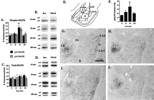
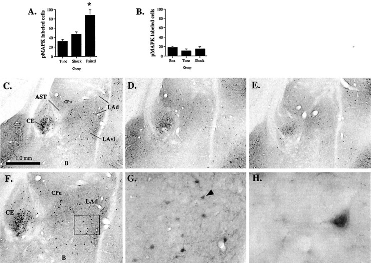
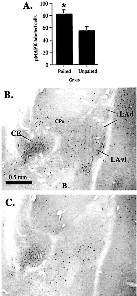
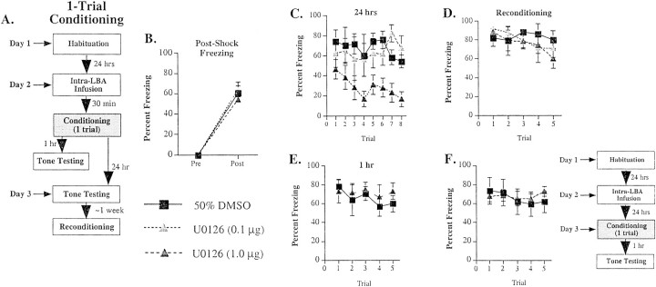
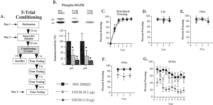
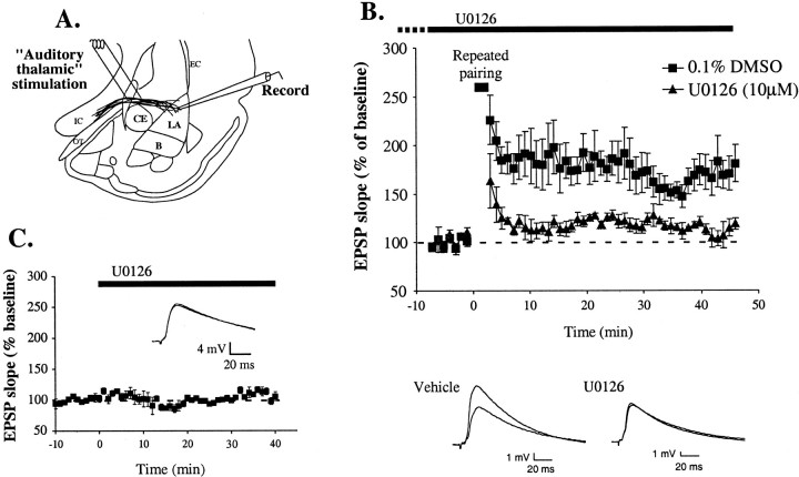
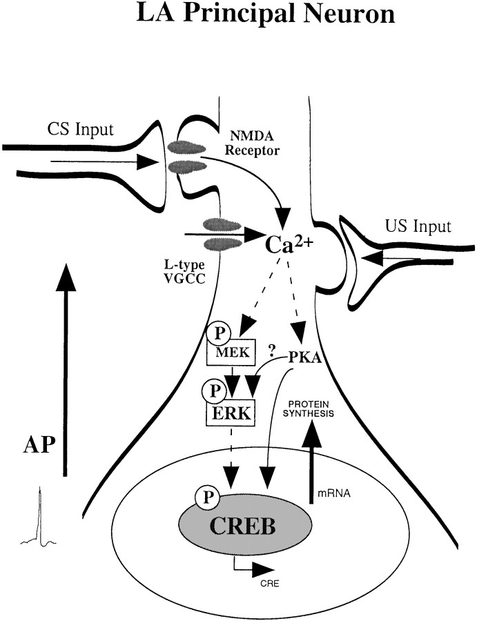
Although much has been learned about the neurobiological mechanisms underlying Pavlovian fear conditioning at the systems and cellular levels, relatively little is known about the molecular mechanisms underlying fear memory consolidation. The present experiments evaluated the role of the extracellular signal-regulated kinase/mitogen-activated protein kinase (ERK/MAPK) signaling cascade in the amygdala during Pavlovian fear conditioning. We first show that ERK/MAPK is transiently activated-phosphorylated in the amygdala, specifically the lateral nucleus (LA), at 60 min, but not 15, 30, or 180 min, after conditioning, and that this activation is attributable to paired presentations of tone and shock rather than to nonassociative auditory stimulation, foot shock sensitization, or unpaired tone-shock presentations. We next show that infusions of U0126, an inhibitor of ERK/MAPK activation, aimed at the LA, dose-dependently impair long-term memory of Pavlovian fear conditioning but leaves short-term memory intact. Finally, we show that bath application of U0126 impairs long-term potentiation in the LA in vitro. Collectively, these results demonstrate that ERK/MAPK activation is necessary for both memory consolidation of Pavlovian fear conditioning and synaptic plasticity in the amygdala.


Figures

Fig. 1.
Fig. 1.
Time course of ERK/MAPK activation in the amygdala. A, Mean ± SE percent pMAPK immunoreactivity from amygdala punches taken from rats decapitated at 15 (n = 10), 30 (n = 10), 60 (n = 14), or 180 (n = 10) min after conditioning. Rats were presented with five tone–foot shock pairings. Paired samples in each group were normalized relative to sham-trained (Box) controls (for details, see Materials and Methods). p42 and p44 correspond to the molecular weights (in kilodaltons) of the two isoforms of mammalian ERK (ERK1 and ERK2) that are recognized by the pMAPK antibody. *p< 0.05 relative to the 15 min time point. B, Representative pMAPK blots from Box andPaired conditions at different time points after conditioning. C, Mean ± SE percent tMAPK immunoreactivity at different time points after conditioning (15, 30, 60, or 180 min) from the samples in A. Paired samples in each group were normalized relative to sham-trained (Box) controls.D, Representative tMAPK blots from Boxand Paired conditions at different time points after conditioning. E, Schematic of the amygdala at approximately bregma −3.3 (according to Paxinos and Watson, 1997). F, Mean ± SE pMAPK-immunoreactive cells in the LA at 15 (n = 5), 30 (n = 5), 60 (n = 5), or 180 (n = 5) min after conditioning. Rats were presented with five tone–foot shock pairings. *p < 0.05 relative to the 15 min time point. G, Representative photomicrograph of pMAPK labeling in the amygdala at 15 min after conditioning. CPu, Caudate/putamen; EC, external capsule. H, Representative photomicrograph of pMAPK labeling in the amygdala at 30 min after conditioning.I, Representative photomicrograph of pMAPK labeling in the amygdala at 60 min after conditioning. J, Representative photomicrograph of pMAPK labeling in the amygdala at 180 min after conditioning.
Fig. 2.
Fig. 2.
Pairing specificity of ERK/MAPK activation in the LA. A, Mean ± SE pMAPK-immunoreactive cells in the LA after presentation of tone alone (Tone;n = 5), foot shock alone (Shock;n = 5), or tone–foot shock pairings (Paired; n = 5). Rats were given five presentations of each stimulus and perfused 60 min later. *p < 0.05 relative to the other groups.B, Mean ± SE pMAPK-immunoreactive cells in the LA after box alone (Box; n = 3), tone alone (Tone; n = 3), or foot shock alone (Shock; n = 3). Rats were perfused 60 min after treatment. C, Representative photomicrograph of pMAPK labeling in the amygdala after conditioning.AST, Amygdala/striatal transition zone.D, Representative photomicrograph of pMAPK labeling in the amygdala after foot shock alone. E, Representative photomicrograph of pMAPK labeling in the amygdala after tone alone.F, 10× magnification of pMAPK labeling in the LA and surrounding nuclei from C. G, 40× magnification of pMAPK labeling in the LA from the insetin F. H, 100× magnification of pMAPK labeling in an LA pyramidal cell (from G;arrow), showing nuclear labeling.
Fig. 3.
Fig. 3.
Associative specificity of ERK/MAPK activation in the LA. A, Mean ± SE pMAPK-immunoreactive cells in the LA after paired (Paired; n = 5) or unpaired (Unpaired; n = 5) presentations of tone and shock. Rats were perfused 60 min later. *p < 0.05 relative to the unpaired group.B, Representative photomicrograph of pMAPK labeling in the amygdala after paired stimulation. C, Representative photomicrograph of pMAPK labeling in the amygdala after unpaired stimulation.
Fig. 4.
Fig. 4.
Effects of intra-LBA administration of U0126 on single-trial fear conditioning. A, Schematic of behavioral protocol. B, Mean ± SE post-shock freezing immediately after the conditioning trial in rats given intra-LBA infusions of 50% DMSO (vehicle; n = 4), 0.1 μg of U0126 (n = 4), or 1.0 μg of U0126 (n = 8). Rats were given a single tone–foot shock pairing. C, Mean ± SE auditory LTM in the rats from B. Rats were assessed for LTM at 24 hr after conditioning. D, Mean ± SE auditory fear memory after reconditioning in the rats from C. Rats were reconditioned drug-free ∼1 week after the initial drug infusions, training, and testing. E, Mean ± SE auditory STM in rats given intra-LBA infusions of 50% DMSO vehicle (n = 8) or 1.0 μg of U0126 (n= 8). Rats were assessed for STM at 1 hr after conditioning.F, Mean ± SE auditory STM in rats given intra-LBA infusions of 50% DMSO vehicle (n = 6) or 1.0 μg of U0126 (n = 6) 24 hr before conditioning and STM testing (see adjacent schematic of behavioral procedures).
Fig. 5.
Fig. 5.
Effects of intra-LBA administration of U0126 on multiple-trial fear conditioning. A, Schematic of behavioral protocol. B, Representative blots and mean ± SE percent pMAPK immunoreactivity from amygdala punches taken from rats given intra-LBA infusions of 50% DMSO (vehicle;n = 6), 0.1 μg of U0126 (n = 6), or 1.0 μg of U0126 (n = 6). *p < 0.05 relative to vehicle controls.C, Mean ± SE post-shock freezing between conditioning trials in rats given intra-LBA infusions of 50% DMSO (vehicle; n = 8), 0.1 μg of U0126 (n = 4), or 1.0 μg of U0126 (n = 7). Rats were given five tone–foot shock pairings. D, Mean ± SE auditory fear memory assessed at 1 hr after conditioning in the rats from C.E, Mean ± SE auditory fear memory assessed at 3 hr after conditioning in the rats from C. F, Mean ± SE auditory fear memory assessed at 6 hr after conditioning in the rats from C. G, Mean ± SE auditory fear memory assessed at 24 hr after conditioning in the rats from C.
Fig. 6.
Fig. 6.
Histological verification of cannula placements.A, Cannula tip placements from rats trained with a single pairing and tested for LTM 24 hr later (see Fig.4B–D). Rats were infused with ACSF (black squares), 0.1 μg of U0126 (white triangles), or 1.0 μg of U0126 (dark gray triangles).B, Cannula tip placements from rats trained with a single pairing and tested for STM 1 hr later (see Fig.4E). Rats were infused with ACSF (black squares) or 1.0 μg of U0126 (dark gray triangles). C, Cannula tip placements from rats trained with a single pairing and tested for STM 1 hr later (see Fig.4F). Rats were infused with ACSF (black squares) or 1.0 μg of U0126 (dark gray triangles) 24 hr before conditioning and STM testing.D, Cannula tip placements from rats trained with multiple pairings and tested for fear memory at 1, 3, 6, and 24 hr after conditioning (see Fig. 5C–G). Rats were infused with ACSF (black squares), 0.1 μg of U0126 (white triangles), or 1.0 μg of U0126 (dark gray triangles). Panels were adapted from Paxinos and Watson (1997).
Fig. 7.
Fig. 7.
Impaired amygdala LTP by U0126. A, Schematic of the amygdala slice preparation, showing placement of stimulating and recording electrodes. Afferent fibers from the auditory thalamus enter the LA medially, coursing through the ventralmost part of the striatum just above the central nucleus. Recordings were made just below the site of termination of auditory thalamic fibers terminating in the LAd. IC, Internal capsule;OT, optic tract; EC, external capsule.B, Mean ± SE percent EPSP slope (relative to baseline) in cells treated with 0.1% DMSO vehicle (n = 5; black squares) or 10 μm U0126 (n = 4; gray triangles) before and after LTP induction. U0126 was applied at the time indicated by solid bar, plus variable time before breaking into the cell indicated by dashed bar.Traces from an individual experiment before and 40 min after induction are shown in the inset.Traces are averages of five responses. C, Mean ± SE percent EPSP slope (relative to baseline) in cells (n = 3) before and after treatment with U0126 (10 μm; solid bar). Traces from an individual experiment before and 30 min after application of U0126 are shown in the inset. Traces are averages of five responses.
Fig. 8.
Fig. 8.
Schematic of the cellular events that may underlie formation of long-term fear memories in the amygdala. Pairing of CS and US inputs in LA principal cells leads to calcium influx through either NMDA receptors or L-type VGCCs. L-type channels are opened on dendritic shafts and spines, possibly by backpropagating action potentials (AP) during training. The increase in intracellular Ca2+ leads to the activation of protein kinases, such as PKA and ERK/MAPK. Once activated, these kinases can translocate to the nucleus where they activate transcription factors such as CREB. The activation of CREB by PKA and ERK/MAPK promotes CRE-mediated gene transcription and the synthesis of new proteins. PKA, ERK/MAPK, CREB, RNA, and protein synthesis in the amygdala have all been shown to be necessary for the establishment of long-term fear memories.

References

    1. Abel T, Nguyen PV, Barad M, Deuel TAS, Kandel ER, Bourchuladze R. Genetic demonstration of a role for PKA in the late phase of LTP and in hippocampus-based long-term memory. Cell. 1997;88:615–626. - PubMed
    1. Atkins CM, Selcher JC, Petraitis JJ, Trzaskos JM, Sweatt JD. The MAPK cascade is required for mammalian associative learning. Nat Neurosci. 1998;1:602–610. - PubMed
    1. Bailey DJ, Kim JJ, Sun W, Thompson RF, Helmstetter FJ. Acquisition of fear conditioning in rats requires the synthesis of mRNA in the amygdala. Behav Neurosci. 1999;113:276–282. - PubMed
    1. Berman DE, Hazvi S, Rosenblum K, Seger R, Dudai Y. Specific and differential activation of mitogen-activated protein kinase cascades by unfamiliar taste in the insular cortex of the behaving rat. J Neurosci. 1998;18:10037–10044. - PMC - PubMed
    1. Blum S, Moore AN, Adams F, Dash PK. A mitogen-activated protein kinase cascade in the CA1/CA2 subfield of the dorsal hippocampus is essential for long-term spatial memory. J Neurosci. 1999;19:3535–3544. - PMC - PubMed

Publication types

MeSH terms