. 2000 Nov 7;97(23):12607-12.
doi: 10.1073/pnas.220302597.

The classical progesterone receptor mediates Xenopus oocyte maturation through a nongenomic mechanism

Affiliations

The classical progesterone receptor mediates Xenopus oocyte maturation through a nongenomic mechanism

M Bayaa et al. Proc Natl Acad Sci U S A. .

Abstract

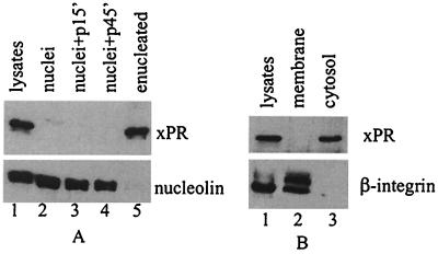
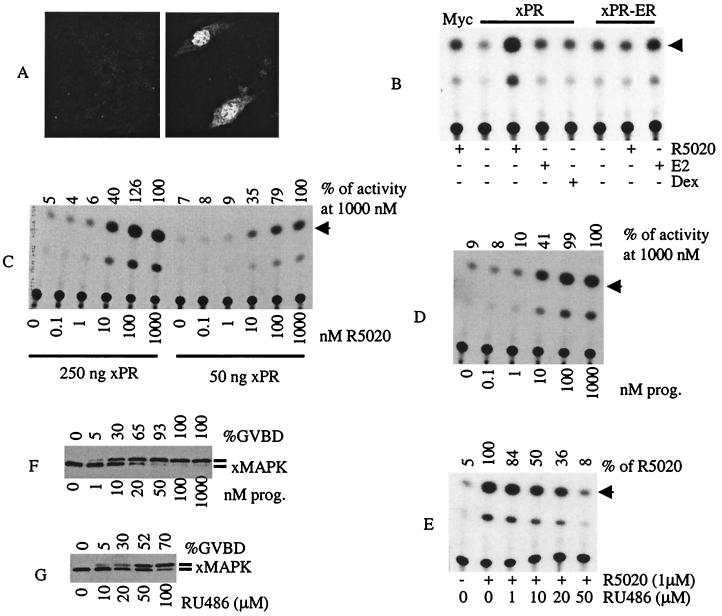
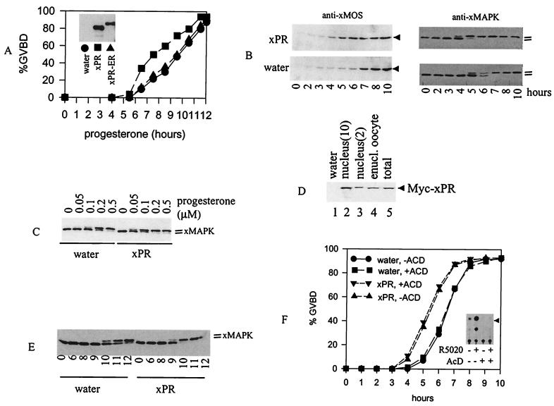
Xenopus laevis oocytes are physiologically arrested at G(2) of meiosis I. Resumption of meiosis, or oocyte maturation, is triggered by progesterone. Progesterone-induced Xenopus oocyte maturation is mediated via an extranuclear receptor and is independent of gene transcription. The identity of this extranuclear oocyte progesterone receptor (PR), however, has remained a longstanding problem. We have isolated the amphibian homologue of human PR from a Xenopus oocyte cDNA library. The cloned Xenopus progesterone receptor (xPR) functioned in heterologous cells as a progesterone-regulated transcription activator. However, endogenous xPR was excluded from the oocyte nucleus and instead appeared to be a cytosolic protein not associated with any membrane structures. Injection of xPR mRNA into Xenopus oocytes accelerated the progesterone-induced oocyte maturation and reduced the required concentrations of progesterone. In enucleated oocytes, xPR accelerated the progesterone-induced mitogen-activated protein kinase activation. These data suggest that xPR is the long sought after Xenopus oocyte receptor responsible for progesterone-induced oocyte maturation.


Figures

Figure 1
Figure 1
Cloning and sequence analyses of xPR. Schematic representation of xPR sequence as compared with human PR sequence (GenBank accession no. M15716). The introns (line) and exons (E1 and E2) are not drawn in proportion in the approximately 9-kb genomic clone. Other numbers indicate amino acid positions. DBD, DNA binding domain; NSL, nuclear localization signal.
Figure 2
Figure 2
Expression of xPR in Xenopus oocytes. (A) Total RNA (1 μg each) from oocytes of the various stages (stages I–III mixed with unknown ratio) were reverse-transcribed followed by PCR amplification using xPR primers encompassing exons E1 and E2. Lane 4 represents a negative control in which PCR was performed directly on input stage VI oocyte RNA without prior reverse transcription (RT). Shown is a representative of four independent experiments with arrows indicating the specifically amplified products. (B) Extracts from stage IV (lane 1), stage V (lane 2), or stage VI (lane 3) oocytes, isolated after collagenase treatment of ovarian tissues, or extracts from collagenase-treated and devitellinated oocytes (lane 4), were immunoblotted with anti-xPR. Equal amounts (50 μg) of proteins were loaded on each lane. The primary antibodies were visualized by incubation with appropriate secondary antibody-horseradish peroxidase conjugates followed by the use of a chemiluminescence kit (ECL, Amersham Pharmacia). Shown is a representative of three independent experiments. (C) Extracts from uninjected (lanes 1, 5, and 9) or Myc-xPR mRNA-injected (lanes 2, 6, and 10) oocytes (each representing half an oocyte), or extracts from untransfected (lanes 3, 7, and 11) or Myc-xPR-transfected (lanes 4, 8, and 12) COS cells (each representing one-tenth of a 3-cm dish) were immunoblotted with the indicated antibodies. Shown is a representative of three independent experiments.
Figure 3
Figure 3
xPR is a cytosolic protein in Xenopus oocytes. (A) Extracts from intact oocytes (lane 1, equivalent of half an oocyte), isolated nuclei (lane 2, 1 GV), nuclei isolated from oocytes after 15- or 45-min incubation with progesterone (lanes 3 and 4, respectively, 1 GV each), or from enucleated oocytes (lane 5, half an oocyte), were immunoblotted with anti-xPR or anti-Xenopus nucleolin (R2D2) (14). Shown is a representative of five independent experiments. (B) Extracts from intact oocytes (lane 1), 100,000 × g pellet (lane 2), or supernatant (lane 3), each of which were the equivalent of one oocyte, were blotted with anti-xPR or anti-β-integrin. Shown is a representative of five independent experiments.
Figure 4
Figure 4
xPR functions as a transcription activator in COS cells. (A) Typical confocal images of mock-transfected (Left) or Myc-xPR transfected COS cells (Right) immunostained with anti-Myc antibodies. (B) COS cells transfected with mouse mammary tumor virus-CAT, together with the indicated plasmid were stimulated with R5020, 17β estradiol (E2), or dexamethasone (Dex). Cell lysates were prepared and subjected to CAT assays. Shown is a representative of three independent experiments. (C) COS cells transfected with the indicated amounts of xPR plasmid were simulated with the indicated concentrations of R5020. Shown is a representative of three independent experiments. (D) Experiments were similar to C except for using progesterone instead of R5020. (E) xPR-transfected COS cells were treated with R5020 (as indicated) or R5020 together with the indicated concentrations of RU486. Shown is a representative of three independent experiments. (F) Freshly isolated oocytes were treated overnight with the indicated concentrations of progesterone. After scoring each sample for GVBD (expressed as % of total treated oocytes), extracts were prepared and analyzed by xMAP kinase immunoblotting. Shown is a representative of three independent experiments. (G) Experiments similar to F except for using RU486 instead of progesterone.
Figure 5
Figure 5
xPR potentiates progesterone-induced MAP kinase activation and GVBD. (A) Oocytes (>60 oocytes per group) injected with water or xPR or xPR-ER mRNA were incubated with 0.5 μM of progesterone. GVBD was scored at the indicated time after the addition of progesterone. Shown is a representative of four independent experiments. (Inset) Typical expression of xPR or xPR-ER in mRNA-injected oocytes as determined via anti-Myc immunoblotting. We estimated that the amount of xPR derived from the injected mRNA was 5–10 times that of endogenous xPR, based on immunoblotting with anti-xPR antibodies (see Fig. 2C). (B) Oocytes injected with water (>200 oocytes) or xPR (>200 oocytes) were treated with progesterone (0.5 μM). At the indicated time, 20–25 oocytes were withdrawn randomly and lysed immediately. All samples were subjected to immunoblotting with anti-xMOS or anti-xMAPK, as indicated. Shown is a representative of three independent experiments. (C) Oocytes injected with water or xPR mRNA were incubated overnight with the indicated concentrations of progesterone and subjected to anti-xMAP kinase immunoblotting. Shown is a representative of three independent experiments. (D) Oocytes injected with water (lane 1) or Myc-xPR mRNA (lanes 2–5) were subjected to nuclear isolation. Extracts from intact oocytes (lanes 1 and 5, one oocyte each), enucleated oocytes (lane 4, one oocyte), or nuclei (lanes 2 and 3, 10 and two nuclei, respectively) were blotted with anti-Myc. Shown is a representative of two independent experiments. (E) Oocytes injected with water (100 oocytes) or xPR (100 oocytes) were individually enucleated (11). The nucleated oocytes were pooled before being divided into six groups of 15 each and treated with progesterone (0.5 μM). At the indicated time after the addition of progesterone, enucleated oocytes were lysed and subjected to immunoblotting with anti-xMAP kinase. Shown is a representative of three independent experiments. (F) Oocytes injected with water, or xPR mRNA, each were split into two groups (>60 oocytes per group) and immediately placed in OR2 or OR2 containing 5 μg/ml of AcD. After a 24-h incubation, progesterone (0.5 μM) was added to all four groups. GVBD was scored at the indicated time after the addition of progesterone. Shown is a representative of two independent experiments. (Inset) CAT assays of xPR-transfected COS cells treated with R5020 (1 μM), AcD (5 μg/ml), alone or in combination.

Comment in

References

    1. Masui Y. J Exp Zool. 1967;166:365–376. - PubMed
    1. Masui Y, Markert C L. J Exp Zool. 1971;177:129–146. - PubMed
    1. Smith L D. Development (Cambridge, UK) 1989;107:685–699. - PubMed
    1. Maller J L. Biol Cell. 1998;90:453–460. - PubMed
    1. Sadler S E, Maller J L. J Steroid Biochem. 1985;22:419–426. - PubMed

Publication types

MeSH terms

Associated data