. 2000 Nov 7;97(23):12571-6.
doi: 10.1073/pnas.220417997.

Formation of high molecular weight complexes of mutant Cu, Zn-superoxide dismutase in a mouse model for familial amyotrophic lateral sclerosis

Affiliations

Formation of high molecular weight complexes of mutant Cu, Zn-superoxide dismutase in a mouse model for familial amyotrophic lateral sclerosis

J A Johnston et al. Proc Natl Acad Sci U S A. .

Abstract

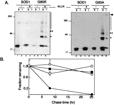
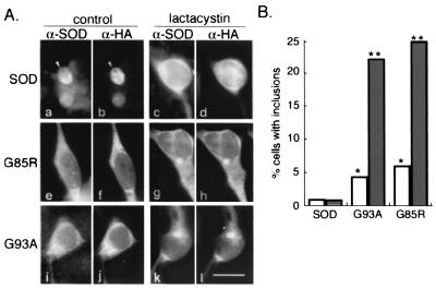
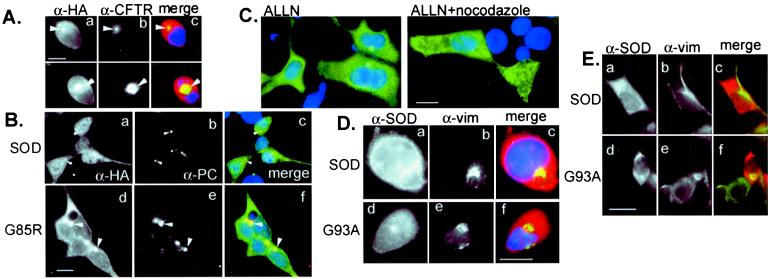
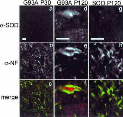
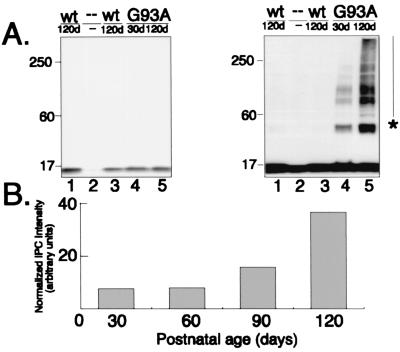
Deposition of aggregated protein into neurofilament-rich cytoplasmic inclusion bodies is a common cytopathological feature of neurodegenerative disease. How-or indeed whether-protein aggregation and inclusion body formation cause neurotoxicity are presently unknown. Here, we show that the capacity of superoxide dismutase (SOD) to aggregate into biochemically distinct, high molecular weight, insoluble protein complexes (IPCs) is a gain of function associated with mutations linked to autosomal dominant familial amyotrophic lateral sclerosis. SOD IPCs are detectable in spinal cord extracts from transgenic mice expressing mutant SOD several months before inclusion bodies and motor neuron pathology are apparent. Sequestration of mutant SOD into cytoplasmic inclusion bodies resembling aggresomes requires retrograde transport on microtubules. These data indicate that aggregation and inclusion body formation are mechanistically and temporally distinct processes.


Figures

Figure 1
Figure 1
IPC formation by mutant SOD. (A) Effect of proteasome inhibition on electrophoretic mobility and detergent solubility of SOD. HEK cells transiently expressing wild-type SOD (lanes 1–4 and 9–12), G85R (lanes 5–8), or G93A (lanes 13–16) mutant SOD were treated overnight with proteasome inhibitor (ALLN, 10 μg/ml), lysed, and separated into nonionic detergent soluble (s) and insoluble (i) fractions and analyzed on immunoblots probed with a mAb to the HA epitope tag. Mobility of molecular weight markers in kDa is indicated at Left. Asterisks denote positions of high molecular SOD at ×2 and ×3 the apparent molecular weight of monomeric SOD. (B) Decreased stability of mutant SOD. HEK cells transiently expressing wild-type (○ and □) or G85R (● and ■) SOD were pulse-labeled with [35S]Cys for 45 min and chased for the times indicated in the presence (□ and ■) or absence (○ and ●) of the proteasome inhibitor lactacystin (5 μM). Data are mean ± SEM from 4–6 independent experiments. Similar data were obtained for the G93A mutant.
Figure 2
Figure 2
Mutant SOD accumulates in intracellular inclusion bodies. (A) HEK cells were transfected wild-type or mutant SOD as indicated. Cells on the Right (c, d, g, h, k, and l) were treated overnight with lactacystin, followed by examination by indirect immunofluorescence with an affinity-purified antibody to SOD (α-SOD; a, c, e, g, i, and k) and a mAb to the HA epitope tag (α-SOD; b, d, f, h, j, and l). (Bar = 15 μm.) (B) Cells transfected and treated as in A were counted and scored for the presence of inclusion bodies. Between 200 and 430 cells were counted for each condition. Single (P < 0.05) and double (P < 0.001) asterisks indicate statistical significance relative to untreated SOD based on Student's t test.
Figure 3
Figure 3
Inclusion bodies containing mutant SOD are aggresomes. (A) Colocalization of G93A SOD with ΔF508 CFTR aggresomes. HEK cells were cotransfected with G93A and ΔF508 CFTR, treated with MG132 before examination by double indirect immunofluorescence for CFTR (α-CFTR; b) and HA epitope tag (α-HA; a). (B) Colocalization of SOD and pericentrin. HEK cells were transfected with wild-type (a–c) or G85R mutant (d–f) SOD, examined by double indirect immunofluorescence by using a polyclonal antibody to pericentrin (α-PC; b and e) and mAb to the HA epitope tag (α-HA; a and d). (C) Delivery of SOD to aggresomes requires an intact microtubule cytoskeleton. HEK cells expressing G85R SOD were exposed to proteasome inhibitor (ALLN, 10 μg/ml) in the presence or absence of nocodazole (10 μg/ml) as indicated. SOD immunolocalization was detected with α-HA mAb. (D) Vimentin colocalizes with mutant but not wild-type SOD. HEK cells expressing wild-type (a–c) or G93A (d–f) SOD were exposed overnight to the proteasome inhibitor MG132, examined by double indirect immunofluorescence by using a polyclonal antibody to SOD (α-SOD; a and c) and mAb to vimentin (α-vim; b and e). (Bar = 15 μm.) (E) Vimentin collapses around SOD aggresomes in the absence of proteasome inhibitor. HEK cells expressing wild-type (a–c) or G93A (d–f) SOD were examined by immunofluorescence with antibodies aginat SOD (a and c) and (b and e). (Bar = 15 μm.)
Figure 4
Figure 4
Inclusion bodies in spinal cords from G93A transgenic mice. Abnormal neurofilament deposition around SOD inclusions in spinal cord motoneurons in G93A transgenic mice at P120 (d–f) but not at P30 (a–c). Morphology of P30 motoneurons is indistinguishable from that of P120 SOD mice (g–i) and from nontransgenic mice (not shown). Spinal cord sections from mice expressing human SOD transgenes were double labeled by indirect immunofluorescence using affinity purified polyclonal antibody to SOD (a, d, and g) and mAb to phosphorylated neurofilament (b, e, and h) and visualized by confocal microscopy. Note magnification in a–d is lower than that in d–i. (Bars = 10 μm.)
Figure 5
Figure 5
Accumulation of SOD in IPCs is an early and specific event in spinal cord from mice expressing G93A human SOD. (A) Expression of SOD in transgenic and control mice. Immunoblots of spinal cord extracts (equal μg total protein per lane) from two different mice expressing a wild-type human SOD transgene (lanes 1 and 3), a G93A human SOD transgene at P30 (lane 4) and P120 (lane 5), and from a nontransgenic littermate (lane 2) probed with affinity-purified B1 polyclonal SOD antibody. Blots on Right and Left differ only in the length of time of exposure of the chemilluminescence reaction to film. (B) SOD IPCs increase with age in G93A spinal cords. An immunoblot similar to that in A was probed with affinity-purified B1 antibody. Intensity of high molecular weight, B1-immunoreactive material was quantified from a digital scan of the blot by integrating the total pixel density after subtraction of background intensity from a nontransgenic control lane.

References

    1. Siddique T. Adv Neurol. 1991;56:227–231. - PubMed
    1. Emery A, Holloway S. In: Human Motor Neuron Diseases. Rowland L, editor. New York: Raven; 1982. pp. 139–147. - PubMed
    1. Rosen D R, Siddique T, Patterson D, Figlewicz D A, Sapp P, Hentati A, Donaldson D, Goto J, O'Regan J P, Deng H X, et al. Nature (London) 1993;362:59–62. - PubMed
    1. Siddique T, Nijhawan D, Hentati A. Neurology. 1996;47:S27–S35. - PubMed
    1. Reaume A G, Elliott J L, Hoffman E K, Kowall N W, Ferrante R J, Siwek D F, Wilcox H M, Flood D G, Beal M F, Brown R H, Jr, et al. Nat Genet. 1996;13:43–47. - PubMed

Publication types

MeSH terms

LinkOut - more resources