. 2000 Nov 7;97(23):12810-5.
doi: 10.1073/pnas.230260397.

Endogenous regulators of G protein signaling proteins regulate presynaptic inhibition at rat hippocampal synapses

Affiliations

Endogenous regulators of G protein signaling proteins regulate presynaptic inhibition at rat hippocampal synapses

H Chen et al. Proc Natl Acad Sci U S A. .

Abstract

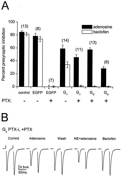
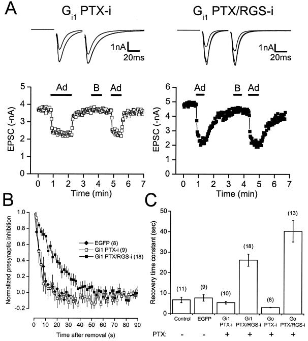
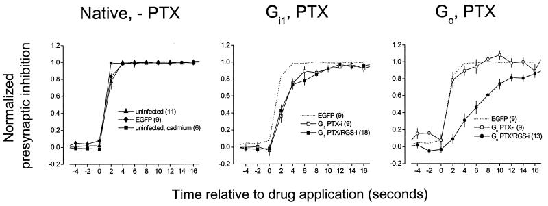
Presynaptic inhibition mediated by G protein-coupled receptors (GPCRs) can develop and decay in a few seconds. This time course is too rapid to be accounted for by the intrinsic GTPase activity of Galpha subunits alone. Here, we test the hypothesis that endogenous regulators of G protein signaling (RGS proteins) are required for rapid, brief presynaptic inhibition. Endogenous G protein alpha subunits were uncoupled from GPCRs by treating cultures with pertussis toxin (PTX). Adenoviral expression of mutant PTX-insensitive (PTX-i) Galpha(i1-3) or Galpha(o) subunits rescued adenosine-induced presynaptic inhibition in cultured hippocampal neurons. Expression of double mutant Galpha(i1) or Galpha(o) subunits that were both PTX-insensitive and unable to bind RGS proteins (PTX/RGS-i) also rescued presynaptic inhibition. Presynaptic inhibition mediated by PTX/RGS-i subunits decayed much more slowly after agonist removal than that mediated by PTX-i subunits or native G proteins. The onset of presynaptic inhibition mediated by PTX/RGS-i Galpha(o) was also slower than that mediated by PTX-i Galpha(o). In contrast, the onset of presynaptic inhibition mediated by PTX/RGS-i Galpha(i1) was similar to that mediated by PTX-i Galpha(i1). These results suggest that endogenous RGS proteins regulate the time course of G protein signaling in mammalian central nervous system presynaptic terminals.


Figures

Figure 1
Figure 1
Rescue of presynaptic inhibition by PTX-insensitive Gαi and Gαo subunits after PTX pretreatment. (A) Summary of experiments comparing presynaptic inhibition induced by baclofen (50 μM) or adenosine (20 μM) under control conditions, with or without infection with adenoviruses expressing various proteins, and with or without pretreatment with PTX (100 ng ml−1, >24 h). Percent presynaptic inhibition was calculated as the difference between peak EPSC amplitudes under control conditions and in the presence of the drug divided by the control EPSC amplitude, multiplied by 100. Bars represent mean ± SEM, with the number of experiments (n) in parentheses. The experimental conditions (virus and toxin treatment) are indicated below each bar. (B) Traces recorded from a neuron expressing PTX-insensitive Gαo after pretreatment with PTX. Adenosine (20 μM) or combined application of adenosine and norepinephrine (NE; 10 μM) depressed evoked EPSCs (with a shift toward paired-pulse facilitation), whereas baclofen (50 μM) was without effect. EPSCs were evoked by pairs of 2-ms depolarizing commands (50-ms interpulse interval) every 2 s (arrows). Each trace represents the average of five consecutive recordings; currents evoked by the depolarizing commands are blanked.
Figure 2
Figure 2
The recovery time course of adenosine-induced presynaptic inhibition mediated by PTX-insensitive and PTX/RGS-insensitive Gα subunits. (A) The time course of presynaptic inhibition is plotted for neurons pretreated with PTX (100 ng ml−1, >24 h) and expressing PTX-insensitive or PTX/RGS-insensitive Gαi1 subunits. Adenosine (20 μM) and baclofen (50 μM) were applied where indicated by the horizontal bars. Note the slow recovery from presynaptic inhibition mediated by PTX/RGS-insensitive Gαi1. Above each plot are superimposed traces (average of five consecutive recordings each) recorded under control conditions, in the presence of adenosine, and in the presence of baclofen. (B) Recovery from presynaptic inhibition (normalized to steady state) is plotted vs. time for neurons expressing EGFP (first 30 s only), PTX-insensitive Gαi1, and PTX/RGS-insensitive Gαi1. Points represent the mean ± SEM; the number of experiments (n) is given in parentheses. (C) Summary of recovery from presynaptic inhibition. Bars represent the mean ± SEM of recovery time constants derived from single exponential fits to plots of EPSC amplitude vs. time after drug removal. The experimental conditions (virus and toxin treatment) are indicated below each bar.
Figure 3
Figure 3
The onset time course of presynaptic inhibition mediated by PTX-insensitive and PTX/RGS-insensitive Gα subunits. Superimposed plots of normalized presynaptic inhibition vs. time, with drug application at 0 s. Each point represents the mean ± SEM, with the number of experiments (n) given in parentheses in the legend. Presynaptic inhibition was normalized by using data points 6 s before and at least 20 s after drug application (20 μM adenosine, 50 μM baclofen). In all experiments where Gα subunits were expressed, neurons were pretreated with PTX.

References

    1. Wu L G, Saggau P. Trends Neurosci. 1997;20:204–212. - PubMed
    1. Thompson S M, Capogna M, Scanziani M. Trends Neurosci. 1993;16:222–227. - PubMed
    1. Ikeda S R, Dunlap K. Adv Second Messenger Phosphoprotein Res. 1999;33:131–151. - PubMed
    1. Wu L G, Saggau P. Neuron. 1994;12:1139–1148. - PubMed
    1. Dittman J S, Regehr W G. J Neurosci. 1996;16:1623–1633. - PMC - PubMed

Publication types

MeSH terms

Substances

LinkOut - more resources