. 2000 Oct 24;97(22):11700-6.
doi: 10.1073/pnas.97.22.11700.

Molecular genetics of pattern formation in the inner ear: do compartment boundaries play a role?

Affiliations

Molecular genetics of pattern formation in the inner ear: do compartment boundaries play a role?

J V Brigande et al. Proc Natl Acad Sci U S A. .

Abstract

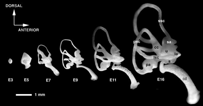
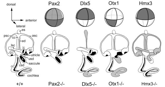
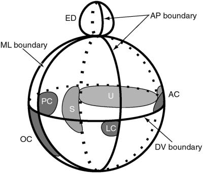
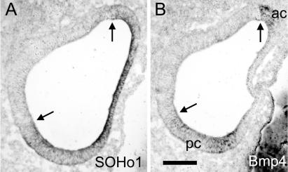
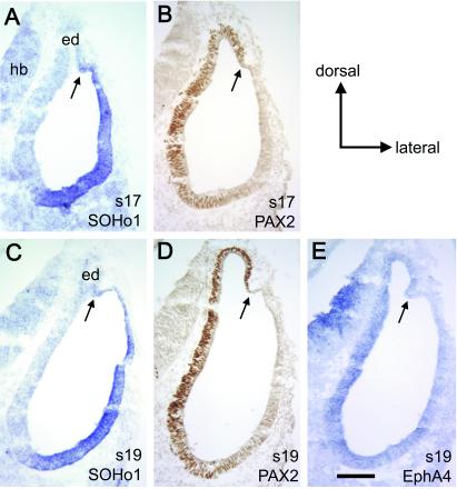
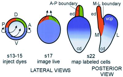
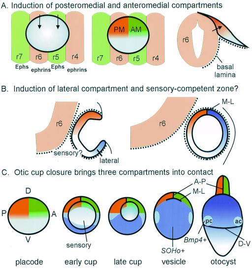
The membranous labyrinth of the inner ear establishes a precise geometrical topology so that it may subserve the functions of hearing and balance. How this geometry arises from a simple ectodermal placode is under active investigation. The placode invaginates to form the otic cup, which deepens before pinching off to form the otic vesicle. By the vesicle stage many genes expressed in the developing ear have assumed broad, asymmetrical expression domains. We have been exploring the possibility that these domains may reflect developmental compartments that are instrumental in specifying the location and identity of different parts of the ear. The boundaries between compartments are proposed to be the site of inductive interactions required for this specification. Our work has shown that sensory organs and the endolymphatic duct each arise near the boundaries of broader gene expression domains, lending support to this idea. A further prediction of the model, that the compartment boundaries will also represent lineage-restriction compartments, is supported in part by fate mapping the otic cup. Our data suggest that two lineage-restriction boundaries intersect at the dorsal pole of the otocyst, a convergence that may be critical for the specification of endolymphatic duct outgrowth. We speculate that the patterning information necessary to establish these two orthogonal boundaries may emanate, in part, from the hindbrain. The compartment boundary model of ear development now needs to be tested through a variety of experimental perturbations, such as the removal of boundaries, the generation of ectopic boundaries, and/or changes in compartment identity.


Figures

Figure 1
Figure 1
Morphogenesis of the chicken inner ear viewed by filling the inner ears with opaque paint. Lateral views are shown. The labyrinth develops from a simple ovoid vesicle. Between embryonic day 3 (E3) and E7, the major structural parts of the ear make their appearance, whereas growth, cell fate specification, and tissue differentiation continue for a longer period. cc, Common crus; la, lateral ampulla; lsc, lateral semicircular canal; psc, posterior semicircular canal; sa, superior ampulla; ssc, superior semicircular canal; u, utricle. Modified from Bissonnette and Fekete (58). Both lateral and posterior views of paint-filled ears have been morphed with Elastic Reality (version 1.3; now owned by Avid) to generate a QuickTime movie that can be found as supplementary material on the PNAS website, www.pnas.org.
Figure 2
Figure 2
Schematic representation of the mouse otic vesicle, showing the expression domains of several genes (top row) and the phenotypes that result when the gene is knocked out (bottom row). Dark lines encircling the vesicle indicate theoretical boundaries that segregate the vesicle into compartments. In the bottom row, structures reported missing in the knockout are left off the drawings, while those that have been reported to have a variable phenotype either by a single group or by two different groups are shaded gray. See text for discussion. Data were derived from the following sources: Pax2 expression, ref. ; pax2−/−, ref. ; Hmx3 expression, refs. and ; Hmx3−/−, refs. and ; Otx1 expression, refs. and ; Otx1−/−, refs. , , and ; Dlx5 expression and Dlx5−/−, ref. . asc, Anterior semicircular canal; ac, anterior crista; cc, common crus; csd, cochlear-saccular duct; ed, endolymphatic duct; es, endolymphatic sac; lc, lateral crista; lsc, lateral semicircular canal; oC, organ of Corti; pc, posterior crista; psc, posterior semicircular canal; s, saccular macula; u, utricular macula; usd, utricular-saccular duct.
Figure 3
Figure 3
Model showing one possible arrangement of sensory primordia with respect to the theoretical compartment boundaries defined in Fig. 2. Both chicken and mouse data were used to derive the model, which is viewed from the side with the same orientation as Fig. 2. Several sensory organ primordia arise on the edge of a SOHo boundary (10). The lateral crista primordium is located on the edge of the Otx2 domain (55). The endolymphatic duct also arises on one side of the medial–lateral (ML) boundary at the dorsal pole of the otocyst (see text and Fig. 5). AC, anterior crista; ED, endolymphatic duct; LC, lateral crista; OC, organ of Corti; PC, posterior crista; S, saccular macula; U, utricular macula.
Figure 4
Figure 4
Sensory organ primordia first arise adjacent to the boundary of a gene, SOHo-1, that identifies a putative lateral compartment. Adjacent horizontal sections through the center of the stage 19 chicken otocyst were probed for SOHo1 mRNA (A) or Bmp4 mRNA (B). At this stage, Bmp4 marks the sensory anlagen of the anterior crista (ac) and the posterior crista (pc) (21). Arrows indicate the places where the two genes appear to share the same boundary. Lateral is to the left and anterior is up. (Scale bar = 100 μm.)
Figure 5
Figure 5
A putative compartment boundary separating medial and lateral gene expression domains can be found near the dorsal pole of the otocyst in the chicken embryo. Adjacent transverse sections through the middle of the chicken otocyst were probed for SOHo1 mRNA (A and C), PAX2 protein (B and D), or EphA4 mRNA (E). The expression of EphA4 is generally weak, but appears to map to the same location as PAX2 at stage 19 (s19), as well as all stages from 18 to 27 (data not shown). Note that all three genes appear to overlap in the ventral but not the dorsal otocyst. Arrows indicate the position of a putative M-L boundary just lateral to the developing endolymphatic duct (ed). (Scale bar = 100 μm.) hb, Hindbrain.
Figure 6
Figure 6
A schematic of the results of fate mapping the chick otic cup. The data suggest that two lineage-restriction boundaries intersect at the dorsal pole of the otocyst. The M-L lineage-restriction boundary corresponds to the same location as the boundary defined by SOHo1 and PAX2 expression domains (compare with Fig. 5). The anterior–posterior (A-P) lineage-restriction boundary appears to bisect the endolymphatic duct (ed). Arrows indicate directions of growth seen at subsequent stages. This schematic is based on data from ref. . D, dorsal; V, ventral; cd, cochlear duct; s, stage; vcp, vertical canal pouch.
Figure 7
Figure 7
A schematic showing possible inductive influences of hindbrain on the development of putative compartments in the otic vesicle. (A) The r5-r6 boundary in the hindbrain is aligned with the A-P lineage-restriction boundary in the otic cup. Thus, r5 and r6 are appropriately positioned to send different inductive signals (arrows) to anterior and posterior otic placode, respectively. Because only the dorsal half of the placode makes intimate contact with the hindbrain cells because of the absence of a basal lamina between the two structures (34), inductive signals that require direct cell contact may not extend to the ventral rim of the otic field, as shown. (B) It is proposed that the ventral tissue does not acquire lateral identity until the cup stage, and is separated from the dorsomedial tissue by a region that acquires sensory competence. The signaling sources for either lateral or sensory identity are unknown, but they could be influenced by distance from the hindbrain and/or proximity to lateral ectoderm or mesoderm. (C) A schematic of otic vesicle formation, combining information from fate mapping and gene expression domains of SOHo and Bmp4. The gray region in the center of the field is proposed to correspond to a sensory-competent region that will intersect with the broader gene expression domains to form the sensory patches and perhaps the ganglion cells. Only the formation of the anterior crista (ac) and posterior crista (pc) is shown. The timing of appearance and location of a putative dorsal–ventral (D-V) boundary, although predicted to be present by stage 22 to place the cristae at the right location, has not yet been confirmed by fate mapping. cd, Cochlear duct; ed, endolymphatic duct; r, rhombomere.

References

    1. Lang H, Bever M M, Fekete D M. J Comp Neurol. 2000;417:205–220. - PubMed
    1. Fekete D M. Curr Opin Neurobiol. 1996;6:533–541. - PubMed
    1. Whitfield T, Haddon C, Lewis J. Semin Cell Dev Biol. 1997;8:239–247. - PubMed
    1. Torres M, Giraldez F. Mech Dev. 1998;71:5–21. - PubMed
    1. Rivolta M N. Audiol Neurootol. 1997;2:36–49. - PubMed

Publication types

LinkOut - more resources