. 2000 Oct 24;97(22):11736-43.
doi: 10.1073/pnas.97.22.11736.

Cochlear mechanisms from a phylogenetic viewpoint

Affiliations

Cochlear mechanisms from a phylogenetic viewpoint

G A Manley. Proc Natl Acad Sci U S A. .

Abstract

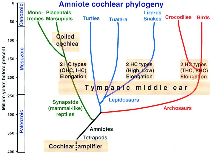
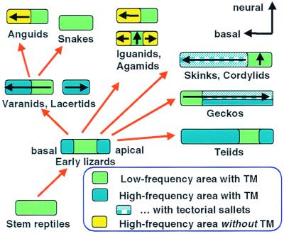
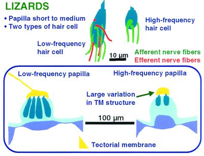
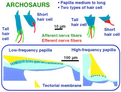
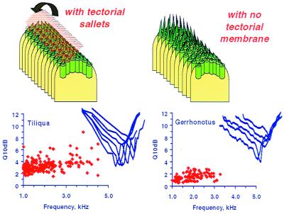
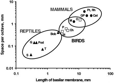
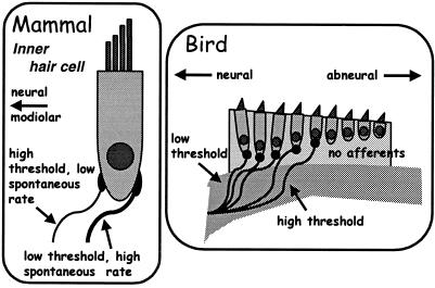
The hearing organ of the inner ear was the last of the paired sense organs of amniotes to undergo formative evolution. As a mechanical sensory organ, the inner-ear hearing organ's function depends highly on its physical structure. Comparative studies suggest that the hearing organ of the earliest amniote vertebrates was small and simple, but possessed hair cells with a cochlear amplifier mechanism, electrical frequency tuning, and incipient micromechanical tuning. The separation of the different groups of amniotes from the stem reptiles occurred relatively early, with the ancestors of the mammals branching off first, approximately 320 million years ago. The evolution of the hearing organ in the three major lines of the descendents of the stem reptiles (e.g., mammals, birds-crocodiles, and lizards-snakes) thus occurred independently over long periods of time. Dramatic and parallel improvements in the middle ear initiated papillar elongation in all lineages, accompanied by increased numbers of sensory cells with enhanced micromechanical tuning and group-specific hair-cell specializations that resulted in unique morphological configurations. This review aims not only to compare structure and function across classification boundaries (the comparative approach), but also to assess how and to what extent fundamental mechanisms were influenced by selection pressures in times past (the phylogenetic viewpoint).


Figures

Figure 1
Figure 1
Highly schematic representation of the amniote phylogenetic tree over 400 million years to illustrate the approximate time of origin of particular features of auditory systems. Amniotes arose from the earliest tetrapods early in the paleozoic and inherited from them a simple hearing organ with a cochlear amplifier in the stereovillar bundles. Apart from the lineages to the turtles and the Tuatara, that remained primitive in a number of respects, three main lineages to modern amniotes are distinguished here. Splitting off first were mammalian ancestors, which gave rise to both the egg-laying monotremes and the marsupial-placental line. Later, the archosaur line originated and led to the dominant land organisms of the mesozoic. Of these, only the crocodile-alligator and bird groups survived to modern times. The last group to split off was the lizards and snakes within the lepidosaurs. The tympanic middle ear originated independently in all groups during the Triassic, initiating the evolution of unique configurations of papillae, with all groups showing papillar elongation and hair-cell specializations. In mammals IHC and OHC, in birds THC and SHC populations developed. In lizards, the populations segregated along frequency lines (low- and high-frequency populations). Because the hair-cell populations in the monotreme and marsupial-placental mammal groups are so similar, they almost certainly arose before these lineages diverged. The same applies to the birds and Crocodilia. In lizards, there are great family-specific variations, suggesting that these hair-cell populations arose soon after the Triassic. Because monotremes do not have a coiled cochlea, coiling almost certainly developed in the marsupial-placental lineage. [Modified after ref. and used with permission of Wiley-VCH Press (Copyright 2000, Wiley).]
Figure 2
Figure 2
Middle-ear sensitivity in representatives of three amniote groups, shown as the velocity of the center of the eardrum (the tip of the malleus or extracolumella) as a function of frequency at 100 dB sound pressure level (SPL). At this sound pressure, air particles have a velocity of 6 mm/sec (dashed line). The Tokay gecko (gray line, ●) is a sensitive lizard (6), the chicken (black line, ▴) represents birds (7), and the guinea pig (gray line, ■) represents mammals (8). The main difference observed is not in sensitivity, but in the upper frequency limits and the high-frequency flanks.
Figure 3
Figure 3
A schematic summary of the structure of the auditory papilla in a primitive amniote, the red-eared turtle. Most of the hair cells are placed over the BM and covered by a thick TM (yellow). There is only one type of hair cell, which is innervated by both afferent and efferent fibers (Right).
Figure 4
Figure 4
(AC) Basic hair-cell structures contributing to micromechanical frequency tuning. In most cases, a tectorial structure covers the hair-cell bundle. A hair cell with a taller stereovillar bundle and larger tectorial cover (A) responds best to much lower frequencies than a hair cell with a shorter bundle and less massive tectorial structure (B). Equivalent frequencies in hair cells without tectorial structures (C) result from much taller bundles. (D) Schematic of the ion channels involved in electrical tuning of hair cells. The flow of ions (mainly K+) through transduction channels depolarizes the cell and activates voltage-sensitive Ca2+ channels in the baso-lateral membrane, raising the Ca2+ concentration [Ca2+] in the cell and activating Ca2+-sensitive K+ channels. This leads to K+ outflow, hyperpolarization, and a new cycle. Channel number, kinetics, and the temperature determine the frequency of oscillation. In turtles, frequencies between 50 and 600 Hz have been measured, but in other amniotes, higher frequencies are reached (see text).
Figure 5
Figure 5
Highly schematic representation of a possible evolutionary sequence of the papillae of modern lizard families. Where known, the direction of the tonotopicity, from low to high frequencies, is shown (arrow). From stem reptile papillae with uniform hair cells, a papilla arose with new, mirror-imaged, micromechanically tuned hair-cell areas at both ends, flanking the low-frequency (green) area. From this, the various papillar configurations of different lizard families and the snakes can be derived as shown, including the reversed tonotopic organization in geckos. Placing similarly formed papillae together does not necessary imply close systematic relationships. In different families, the TM over the high-frequency areas was either retained (uniformly blue areas), divided up into sallets (patterned blue areas), or lost (yellow areas) (see text; partly after ref. 10). [Modified after ref. , and used with permission of Wiley-VCH Press (Copyright 2000, Wiley).]
Figure 6
Figure 6
A schematic summary of the structure of the auditory papilla of lizards, which always have a low- and a high-frequency area. Over the latter, the tectorial structure (yellow) is highly variable between and even within families and is missing in some groups (see Fig. 5). Low- and high-frequency hair cells differ both in their size and their innervation pattern; high-frequency hair cells (Upper Right) never receive an efferent innervation.
Figure 7
Figure 7
A schematic summary of the structure of the auditory papilla of birds, which are between 2 and 11 mm in length and contain between 3,000 and 17,000 hair cells. The hair cells form a continuum, with the tallest cells at the apical end (Left, transverse section and typical hair-cell shape), where there are many hair cells across the papilla. Hair cells become shorter toward the base, where the number of hair cells across the papilla is much smaller (Right, transverse section and typical hair-cell shapes), and toward the abneural side. All hair cells are covered by a wedge-shaped TM (yellow). Short hair cells have no afferent innervation.
Figure 8
Figure 8
The effect of TM coupling on the responses of lizard papillae. (Left) The salletal TM is shown lifted, to reveal hair-cell bundles. In the alligator lizard Gerrhonotus (Right), the high-frequency papillar area contains free-standing hair cells that lack a TM over their bundles. The requisite frequency range is attained by a strong gradient in bundle height along the papilla. Here, tuning sharpness of afferent fibers is poor (given as the Q10dB of tuning curves—the center frequency divided by the tuning bandwidth at 10 dB above the best sensitivity). (Inset) Representative tuning curves are shown. Where hair cells are attached to tectorial sallets (Left, represented by the Australian Bobtail lizard Tiliqua), the coupling of neighboring hair cells leads to greater sensitivity and higher frequency selectivity, manifest as greater Q10dB values. In addition, the tuning curve flanks in Tiliqua are steeper (6).
Figure 9
Figure 9
The length of the BM and thus of the auditory organ correlates with the space constant for frequency (averaged along the papilla) in reptiles (▴), birds (□), and mammals (●). Because their hearing range did not increase in proportion to the length of the cochlea, avian and mammalian papillae gained much more space for the analysis of any given octave. The increase was greater in most mammals than in most birds, but the distributions overlap. The resulting increase in hair cells and nerve fibers per octave increased parallel processing. Lizards: G, alligator lizard, Gerrhonotus; T, turtle; Pod, European lizards of the genus Podarcis; S, the granite spiny lizard Sceloporus; Bob, Australian Bobtail lizard Tiliqua. Birds: St, starling, Sturnus; Ch, chicken, Gallus; P, pigeon, Columba; O, barn owl, Tyto. Mammals: M, mouse, Mus; GP, guinea pig, Cavia; Cat, house cat, Felis; Pt, Rh, two bat species, Pteronotus, the mustached bat, and Rhinolophus, the horseshoe bat. For refs see also refs. , , and . [Modified after ref. , and used with permission of Wiley-VCH Press (Copyright 2000, Wiley).]
Figure 10
Figure 10
Innervational patterns of primary afferents in mammals and birds that encode auditory sensitivity partly through the specialization of afferent fibers. In mammals (Left), the most sensitive fibers, which also have a higher spontaneous activity, are thicker and innervate the outer side of the IHC. Less sensitive fibers, which have lower spontaneous rates, innervate the modiolar side of the IHC (38). In the starling (Right), different sensitivities of afferent fibers contact different hair cells. The more sensitive fibers contact hair cells near the neural edge, less sensitive fibers hair cells near the middle of the papilla (e.g., ref. 39). Hair cells lying further abneurally have no afferent innervation (28). [Modified after ref. , and used with permission of Wiley-VCH Press (Copyright 2000, Wiley).]

References

    1. Nielsen C. Animal Evolution. Oxford: Oxford Univ. Press; 1995.
    1. Manley G A, Köppl C. Curr Opin Neurobiol. 1998;8:468–474. - PubMed
    1. Clack J A. Brain Behav Evol. 1997;50:198–212. - PubMed
    1. Carr C. In: The Evolutionary Biology of Hearing. Fay R R, Popper A N, Webster D B, editors. New York: Springer; 1992. pp. 511–543.
    1. Manley G A. In: Auditory Worlds: Sensory Analysis and Perception in Animals and Man. Manley G A, Fastl H, Kössl M, Oeckinghaus H, Klump G M, editors. Weinheim: Wiley; 2000. pp. 7–17.

Publication types