. 2000 Oct 24;97(22):11759-64.
doi: 10.1073/pnas.97.22.11759.

Molecular mechanisms of sound amplification in the mammalian cochlea

Affiliations

Molecular mechanisms of sound amplification in the mammalian cochlea

J F Ashmore et al. Proc Natl Acad Sci U S A. .

Abstract

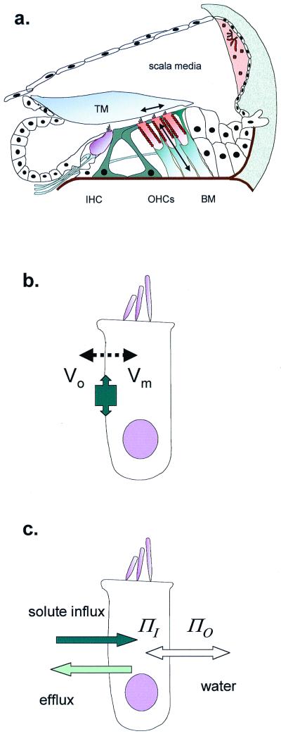
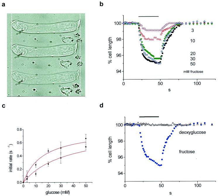
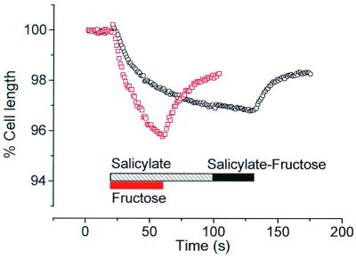
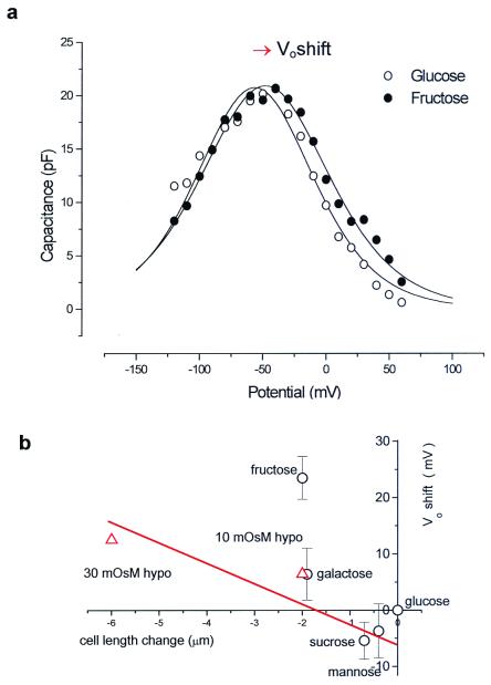
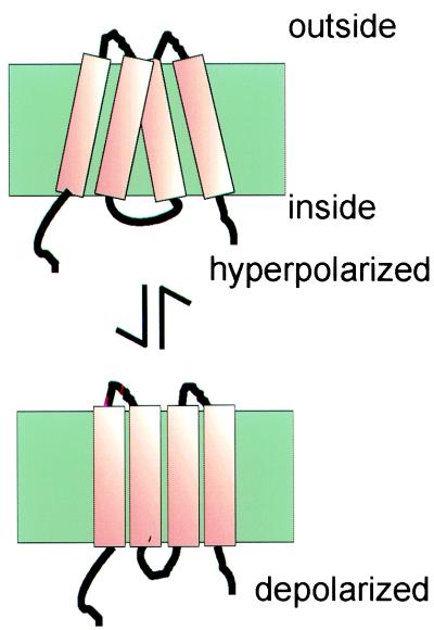
Mammalian hearing depends on the enhanced mechanical properties of the basilar membrane within the cochlear duct. The enhancement arises through the action of outer hair cells that act like force generators within the organ of Corti. Simple considerations show that underlying mechanism of somatic motility depends on local area changes within the lateral membrane of the cell. The molecular basis for this phenomenon is a dense array of particles that are inserted into the basolateral membrane and that are capable of sensing membrane potential field. We show here that outer hair cells selectively take up fructose, at rates high enough to suggest that a sugar transporter may be part of the motor complex. The relation of these findings to a recent candidate for the molecular motor is also discussed.


Figures

Figure 1
Figure 1
OHCs in the cochlea. (a) Schematic cross section of the organ of Corti, showing site of inner hair cells (IHC) and OHCs. The primary stimulus is the shear delivered to the OHC stereocilia by the tectorial membrane (TM). OHC length changes (and therefore forces) are produced as arrowed. BM, basilar membrane. (b) OHC length change through electromotility, where membrane potential (VoVm) alters cell surface area. The tight molecular packing in the lateral membrane allows the protein area change to have macroscopic effects. (c) OHC cell length change through cell volume change, where osmotic pressure (ΠO − ΠI) difference inside and outside requires water to follow solute entry.
Figure 2
Figure 2
Glucose and fructose uptake by guinea-pig OHCs. (a) Fructose-induced cell shortening. Top, control; middle, with fructose; bottom, washout. Cell length, 55 μm. (b) Strains at these different concentrations. Sugars were applied at 3 (n = 6), 10 (n = 7), 20 (n = 8), 30 (n = 6), and 50 mM (n = 7). (c) Initial transport rate estimated by a linear fit between t = 0 s and t = 6 s of glucose (●) or fructose (■) application. Data were fitted with Vmax = 0.8% s−1 and Km = 15.8 ± 1.6 mM (fructose) and 34 ± 3.5 mM (glucose). (d) Effect of 30 mM deoxyglucose (▵) compared with 30 mM fructose (●) shortening. Stimulus timing is shown as a bar in b and d.
Figure 3
Figure 3
Salicylate blocks fructose uptake. Pretreatment of an OHC with 10 mM sodium salicylate induced noticeable cell swelling (○, black) with a stabilization after 80 s. Application of a fructose–salicylate solution on such a pretreated cell failed to induce any response. After washout, the same cell treated with fructose demonstrated a fast and large change in length (□, red). A cell length of 62 μm at the beginning of the experiment is taken as the reference for both recordings.
Figure 4
Figure 4
OHC charge movement is modified by sugars. (a) Membrane capacitance determined by using a staircase ramp protocol. The maximum capacitance Cmax occurs at Vo. In this fit β = 0.036 mV−1. (b) Collected data of Vo shift vs. length change for several sugars (○). Cell capacitance was initially recorded in 330 mosmol⋅kg−1 glucose. Hypoosmotic solution (▵) also produced a Vo shift and length change. The effect of fructose shown in this plot was significantly different from that of other treatments.
Figure 5
Figure 5
Hypothetical model for a motor structure in the OHC membrane. Only four of the transmembrane helices are shown. The net area change in the plane of the membrane, produced by helix tilt, can be sufficient to produce the 5% length change observed in an OHC when electrically stimulated.

References

    1. Manley G A. Proc Natl Acad Sci USA. 2000;97:11736–11743. - PMC - PubMed
    1. Gold T. Proc R Soc London Ser B. 1948;135:492–498.
    1. Flock A, Cheung H. J Cell Biol. 1977;75:339–343. - PMC - PubMed
    1. Hudspeth A J, Choe Y, Mehta A D, Martin P. Proc Natl Acad Sci USA. 2000;97:11765–11772. - PMC - PubMed
    1. Zweig G. J Acoust Soc Am. 1991;89:1229–1254. - PubMed

Publication types

LinkOut - more resources