. 2000 Oct 24;97(22):11773-9.
doi: 10.1073/pnas.97.22.11773.

Detection of synchrony in the activity of auditory nerve fibers by octopus cells of the mammalian cochlear nucleus

Affiliations

Detection of synchrony in the activity of auditory nerve fibers by octopus cells of the mammalian cochlear nucleus

D Oertel et al. Proc Natl Acad Sci U S A. .

Abstract

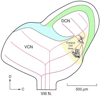
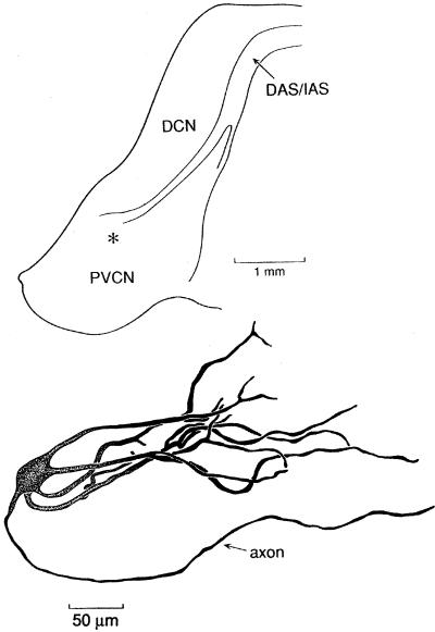
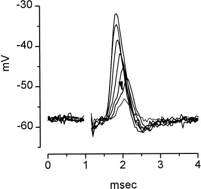
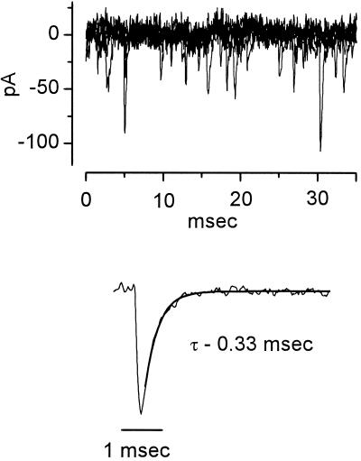
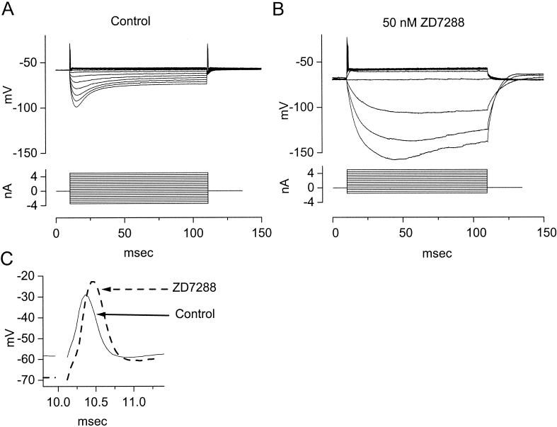
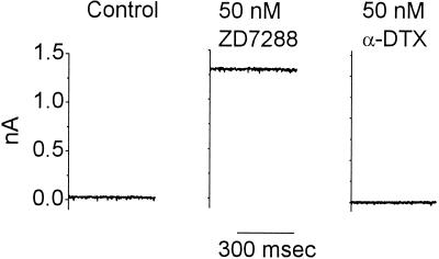
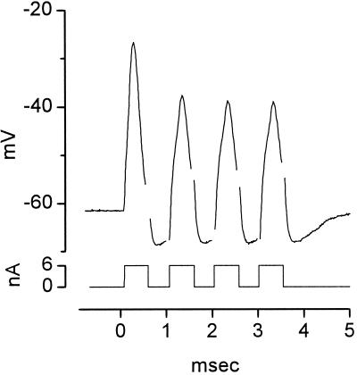
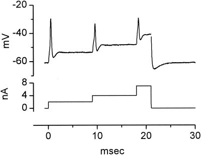
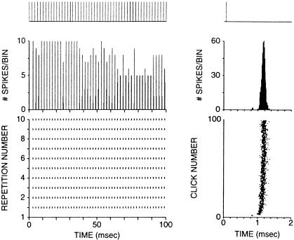
The anatomical and biophysical specializations of octopus cells allow them to detect the coincident firing of groups of auditory nerve fibers and to convey the precise timing of that coincidence to their targets. Octopus cells occupy a sharply defined region of the most caudal and dorsal part of the mammalian ventral cochlear nucleus. The dendrites of octopus cells cross the bundle of auditory nerve fibers just proximal to where the fibers leave the ventral and enter the dorsal cochlear nucleus, each octopus cell spanning about one-third of the tonotopic array. Octopus cells are excited by auditory nerve fibers through the activation of rapid, calcium-permeable, alpha-amino-3-hydroxy-5-methyl-4-isoxazole-propionate receptors. Synaptic responses are shaped by the unusual biophysical characteristics of octopus cells. Octopus cells have very low input resistances (about 7 M Omega), and short time constants (about 200 microsec) as a consequence of the activation at rest of a hyperpolarization-activated mixed-cation conductance and a low-threshold, depolarization-activated potassium conductance. The low input resistance causes rapid synaptic currents to generate rapid and small synaptic potentials. Summation of small synaptic potentials from many fibers is required to bring an octopus cell to threshold. Not only does the low input resistance make individual excitatory postsynaptic potentials brief so that they must be generated within 1 msec to sum but also the voltage-sensitive conductances of octopus cells prevent firing if the activation of auditory nerve inputs is not sufficiently synchronous and depolarization is not sufficiently rapid. In vivo in cats, octopus cells can fire rapidly and respond with exceptionally well-timed action potentials to periodic, broadband sounds such as clicks. Thus both the anatomical specializations and the biophysical specializations make octopus cells detectors of the coincident firing of their auditory nerve fiber inputs.


Figures

Figure 1
Figure 1
Anatomical reconstructions of cell body and dendrites of two intracellularly labeled octopus cells from mice are shown on a schematic version of the cochlear nuclear complex in the parasagittal plane. The granule cell lamina (blue) separates the unlayered VCN from the layered dorsal cochlear nucleus (DCN). Octopus cells occupy an area (yellow) at the most caudal and dorsal extreme of the VCN where auditory nerve fibers are closely bundled as they cross from the VCN to the DCN. Auditory nerve fibers that encode high frequencies (light brown) terminate rostrally and those that encode low frequencies (dark brown) terminate caudally in the octopus cell area. The dendrites of octopus cells extend rostrally from the cell body. Adapted from the results of Golding et al. (19).
Figure 2
Figure 2
Reconstruction with a camera lucida of an octopus cell in a cat that was labeled by an intraxonal injection. The location of the octopus cell body in the coronal section of the cochlear nuclear complex with respect to the posteroventral cochlear nucleus (PVCN), dorsal cochlear nucleus (DCN), dorsal acoustic stria (DAS), and intermediate acoustic stria (IAS) is indicated by * (Upper).
Figure 3
Figure 3
Octopus cells fire in response to the coincident activation of many, but not necessarily all, of the auditory nerve fibers by which they are innervated. Seven superimposed responses are shown to shocks of the auditory nerve of 0.1-msec duration and of varying strength (1–10 V) delivered through a pair of tungsten electrodes. Responses were recorded with a sharp microelectrode filled with 4 M potassium acetate from an octopus cell in a parasagittal slice from the cochlear nucleus of a mouse. The extracellular saline was saturated with 95% oxygen/5% carbon dioxide and contained 130 mM NaCl, 3 mM KCl, 1.3 mM MgSO4, 2.4 mM CaCl2, 20 mM NaHCO3, 3 mM Hepes, 10 mM glucose, 1.2 mM KH2PO4, pH 7.4. Shocks produced artifacts that serve as markers of their occurrence and whose removal left a blank space in the traces. The amplitude of responses was a monotonic function of the shock strength with the weakest shocks producing the smallest responses and the strongest shocks producing the largest responses. The appearance of a small action potential, whose inflection point is marked with an arrowhead, shows where the response was just large and rapid enough to cause firing in the octopus cell. In larger responses the action potential and synaptic potential cannot be resolved. The recording was made by N. L. Golding (19).
Figure 4
Figure 4
Spontaneous miniature synaptic currents recorded from an octopus cell of a mouse under voltage clamp. Patch recording from an octopus cell was made in the whole-cell configuration by using a potassium gluconate-filled pipette. (Upper) Five traces are superimposed to illustrate the frequent spontaneous events when the cell was held near its resting potential at −65 mV. (Lower) Ensemble average of 113 events in the same cell. The decay of currents was well fit with a single exponential with a time constant (τ) equal to 0.33 msec. The pipette solution contained 108 mM potassium gluconate, 9 mM Hepes, 9 mM EGTA, 4.5 mM MgCl2, 14 mM phosphocreatinine (Tris salt), 4 mM ATP (Na salt), and 0.3 mM GTP (Tris salt); pH was adjusted to 7.4 with KOH. The composition of the extracellular saline is given in the legend to Fig. 3. The results have been corrected for a junction potential of −12 mV.
Figure 5
Figure 5
(A) Polarization of an octopus cell with current pulses (−3.5 to 5 nA in 0.5-nA steps) reveals the biophysical characteristics of the cell. Depolarizing current pulses greater than 1 nA produced small action potentials at the onset of the depolarization. After the single action potential, the octopus cell remained depolarized by a few mV. Hyperpolarizing current pulses produced transient hyperpolarizations that sagged back toward rest. (B) In the presence of 50 nM ZD7288, a blocker of gh, the cell hyperpolarized and the input resistance in the hyperpolarizing voltage range increased. The increase in input resistance is reflected in that the current pulses produced larger and slower hyperpolarizations. The rectification in the depolarizing voltage range, reflected in the cluster of traces in responses to depolarizing currents, is not affected by ZD7288. (C) Blocking gh shapes responses in the physiological voltage range. Expansions of the onset of responses to the largest depolarizations shown in A and B show that action potentials are taller and broader in the absence of gh. The voltage drop across the resistance of the electrode was balanced off-line. Whole-cell patch recording from an octopus cell with solutions as in Fig. 4.
Figure 6
Figure 6
At the resting potential Ih is roughly balanced by IK(L). The murine octopus cell was held at its resting potential, −63 mV, under voltage clamp with whole-cell patch clamp under conditions such as those described for Fig. 4. In the presence of ZD7288, a large, steady, outward current developed. Application of α-dendrotoxin blocked a current exactly equal to that which had developed in the presence of ZD7288.
Figure 7
Figure 7
Octopus cells can fire rapidly. A train of depolarizing current pulses presented at 1,000/sec evoked action potentials at every pulse. The voltage drop across the resistance of the electrode was balanced off-line; removal of transient artifacts left brief gaps in the trace. Whole-cell patch recording from an octopus cell of a mouse with solutions as in Fig. 4.
Figure 8
Figure 8
When steadily depolarized, octopus cells are in a relative, but not absolute, refractory period. This octopus cell was depolarized with increasing current steps. Depolarization with a current pulse of 2 nA produced an action potential. Further step depolarizations from 2 to 4 nA and from 4 to 7 nA caused the octopus cell to fire again. Action potentials that were evoked by superimposed step depolarizations were smaller than the initial action potential. At the offset of the current pulse, the octopus cell undershot the resting potential. The voltage drop across the resistance of the electrode was balanced off-line and transient artifacts were made blank. Whole-cell patch recording from a murine octopus cell with solutions as in Fig. 4.
Figure 9
Figure 9
Response of an octopus cell to clicks in a cat in vivo. (Left) Responses to 10 repetitions of a train of acoustic clicks (20-μsec duration) spaced at 2-msec (500 Hz) intervals. (Top) Trace shows the timing of the click stimuli that were presented 30 dB above threshold, (Middle) the poststimulus time histogram (0.2-msec binwidth), and (Bottom) dot rasters to 10 click trains with each symbol representing one action potential. (Right) Responses on an expanded time scale as a function of the 2-msec period of the stimulus. (Top) Trace shows position of the click in the period, (Middle) the period histogram using 8-μsec binwidths, and (Bottom) dot rasters ordered by click number (response to first click on bottom, response to 100th click on top). The threshold of responses to tones at the characteristic frequency, 9 kHz was 52 dB sound pressure level.

References

    1. Joris P X, Smith P H, Yin T C. Neuron. 1998;21:1235–1238. - PubMed
    1. Oertel D. Annu Rev Physiol. 1999;61:497–519. - PubMed
    1. Spirou G A, Davis K A, Nelken I, Young E D. J Neurophysiol. 1999;82:648–663. - PubMed
    1. Takahashi T, Moiseff A, Konishi M. J Neurosci. 1984;4:1781–1786. - PMC - PubMed
    1. Young E D, Sachs M B. J Acoust Soc Am. 1979;66:1381–1403. - PubMed

Publication types

LinkOut - more resources