. 2000 Oct 24;97(22):11793-9.
doi: 10.1073/pnas.97.22.11793.

Subdivisions of auditory cortex and processing streams in primates

Affiliations

Subdivisions of auditory cortex and processing streams in primates

J H Kaas et al. Proc Natl Acad Sci U S A. .

Abstract

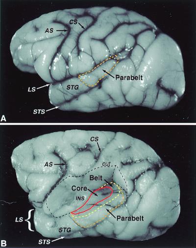
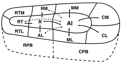
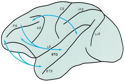
The auditory system of monkeys includes a large number of interconnected subcortical nuclei and cortical areas. At subcortical levels, the structural components of the auditory system of monkeys resemble those of nonprimates, but the organization at cortical levels is different. In monkeys, the ventral nucleus of the medial geniculate complex projects in parallel to a core of three primary-like auditory areas, AI, R, and RT, constituting the first stage of cortical processing. These areas interconnect and project to the homotopic and other locations in the opposite cerebral hemisphere and to a surrounding array of eight proposed belt areas as a second stage of cortical processing. The belt areas in turn project in overlapping patterns to a lateral parabelt region with at least rostral and caudal subdivisions as a third stage of cortical processing. The divisions of the parabelt distribute to adjoining auditory and multimodal regions of the temporal lobe and to four functionally distinct regions of the frontal lobe. Histochemically, chimpanzees and humans have an auditory core that closely resembles that of monkeys. The challenge for future researchers is to understand how this complex system in monkeys analyzes and utilizes auditory information.


Figures

Figure 1
Figure 1
Cortical and subcortical connections of the primate auditory system. Major cortical and subcortical regions are color coded. Subdivisions within a region have the same color. Solid black lines denote established connections. Dashed lines indicate proposed connections based on findings in other mammals. Joined lines ending in brackets denotes connections with all fields in that region. The belt region may include an additional field, MM (see Fig. 5). Abbreviations of subcortical nuclei: AVCN, anteroventral cochlear nucleus; PVCN, posteroventral cochlear nucleus; DCN, dorsal cochlear nucleus; LSO, lateral superior olivary nucleus; MSO, medial superior olivary nucleus; MNTB, medial nucleus of the trapezoid body; DNLL, dorsal nucleus of the lateral lemniscus; VNLL, ventral nucleus of the lateral lemniscus; ICc, central nucleus of the inferior colliculus; ICp, pericentral nucleus of the inferior colliculus; ICdc, dorsal cortex of the inferior colliculus; ICx, external nucleus of the inferior colliculus; MGv, ventral nucleus of the medial geniculate complex; MGd, dorsal nucleus of the medial geniculate complex; MGm, medial/magnocellular nucleus of the medial geniculate complex; Sg, suprageniculate nucleus; Lim, limitans nucleus; PM, medial pulvinar nucleus. Abbreviations of cortical areas: AI, auditory area I; R, rostral area; RT; rostrotemporal area; CL, caudolateral area; CM, caudomedial area; ML, middle lateral area; RM, rostromedial area; AL, anterolateral area; RTL, lateral rostrotemporal area; RTM, medial rostrotemporal area; CPB, caudal parabelt; RPB, rostral parabelt; Tpt, temporoparietal area; TS1,2, superior temporal areas 1 and 2. Frontal lobe areas numbered after the tradition of Brodmann and based on the parcellation of Preuss and Goldmann-Rakic (1): 8a, periarcuate; 46d, dorsal principal sulcus; 12vl, ventrolateral area; 10, frontal pole; orb, orbitofrontal areas.
Figure 2
Figure 2
Lateral view of the macaque cerebral cortex. (A) The approximate location of the parabelt region on the superior temporal gyrus (dashed orange line). (B) Dorsolateral view of the same brain as in A after removal of the overlying parietal cortex, exposing the ventral bank of the lateral sulcus and insula. The approximate locations of the core region (solid red line), caudal and lateral portions of the belt region (dashed yellow line), and the parabelt region (dashed orange line) are shown. The medial portion of the belt region and the rostral pole of the core in the ventral circular sulcus are not visible. Dashed black line defines portion of cortex cut away. AS, arcuate sulcus; CS central sulcus; INS, insula; LS, lateral sulcus; STG superior temporal gyrus; STS, superior temporal sulcus. Adapted from ref. .
Figure 3
Figure 3
Tonotopic organization in the core. Auditory core fields (AI, R, RT) are surrounded by belt fields (not labeled). Curved lines within each field depict isofrequency contours. High- (H) frequency acoustic stimuli are represented caudomedially in AI, rostromedially in R. Low- (L) frequency stimuli are represented rostrolaterally in AI, caudolaterally in R. Tonotopic organization in RT is not as certain but may mirror that found in R. See Fig. 1 for abbreviations.
Figure 4
Figure 4
Architectonic fields in auditory cortex. Macaque brain section flattened and cut parallel to pial surface at 40 μm. Parvalbumin immunohistochemistry. The core fields are the most darkly stained. The caudal belt fields (ML, CL, CM) are moderately dark. Scale bar = 5 mm. Adapted from ref. . See Fig. 1 for abbreviations.
Figure 5
Figure 5
Auditory cortical connections of AI. Area AI, as well as other core areas, has dense reciprocal connections with adjacent areas of the core and belt (solid lines with arrows). Connections with nonadjacent fields are less dense (dashed lines with arrows). The core has few, if any, connections with the parabelt or more distant cortex. See Fig. 1 for abbreviations.
Figure 6
Figure 6
Architectonic identification of core and belt regions. Coronal sections showing the border between the core and belt regions of auditory cortex (arrowheads). Acetylcholinesterase histochemistry. (A) Macaque monkey; (B) chimpanzee; (C) human. Compared with the belt region, the density of acetylcholinesterase in the middle cortical layers (IIIc and IV) is particularly high in the core.
Figure 7
Figure 7
Auditory cortical connections of ML. Area ML, and other belt areas, have dense connections with adjacent areas of the core, belt, and parabelt (solid lines with arrows). Connections with nonadjacent fields tend to be less dense (dashed lines with arrows). The belt fields also have topographically organized connections with functionally distinct fields in prefrontal cortex. Abbreviations defined in Fig. 1.
Figure 8
Figure 8
Auditory cortical connections of CPB. Parabelt area CPB, as well as RPB, has dense connections with adjacent areas of the belt and RM in the medial belt (solid lines with arrows). Connections with nonadjacent fields of the belt tend to be less dense (dashed lines with arrows). The parabelt fields have few, if any, connections with the core areas. The parabelt also has connections with the polysensory areas in the superior temporal sulcus (STS) and with functionally distinct fields in prefrontal cortex. Abbreviations defined in Fig. 1.
Figure 9
Figure 9
Prefrontal connections of auditory cortex in macaque monkeys. Arrows summarize the topographic connections of the lateral belt and parabelt auditory regions with functionally distinct areas of prefrontal cortex. The targets of caudal auditory fields favor spatial domains of prefrontal cortex (e.g., 8a, caudal 46d), whereas more rostral fields exhibit stronger connections with nonspatial regions (e.g., areas 12vl, 10, mediofrontal, orbitofrontal). Connections with intermediate temporal fields tend to overlap more. See Fig. 1 and ref. for more detailed connections. Adapted from ref. , with permission from S. Karger A, Basel.

References

    1. Preuss T M, Goldman-Rakic P S. J Comp Neurol. 1991;310:429–474. - PubMed
    1. Kaas J H, Krubitzer L A. In: Neuroanatomy of Visual Pathways and Their Retinotopic Organization. Dreher B, Robinson S R, editors. London: MacMillan; 1991. pp. 302–359.
    1. Walzl E M. Laryngoscope. 1947;57:778–787. - PubMed
    1. Hackett T A, Stepniewska I, Kaas J H. Brain Res. 1999;817:45–58. - PubMed
    1. Hackett T A, Stepniewska I, Kaas J H. J Comp Neurol. 1998;394:475–495. - PubMed

LinkOut - more resources