. 2000 Oct 24;97(22):11829-35.
doi: 10.1073/pnas.97.22.11829.

Spatial processing in the auditory cortex of the macaque monkey

Affiliations

Spatial processing in the auditory cortex of the macaque monkey

G H Recanzone. Proc Natl Acad Sci U S A. .

Abstract

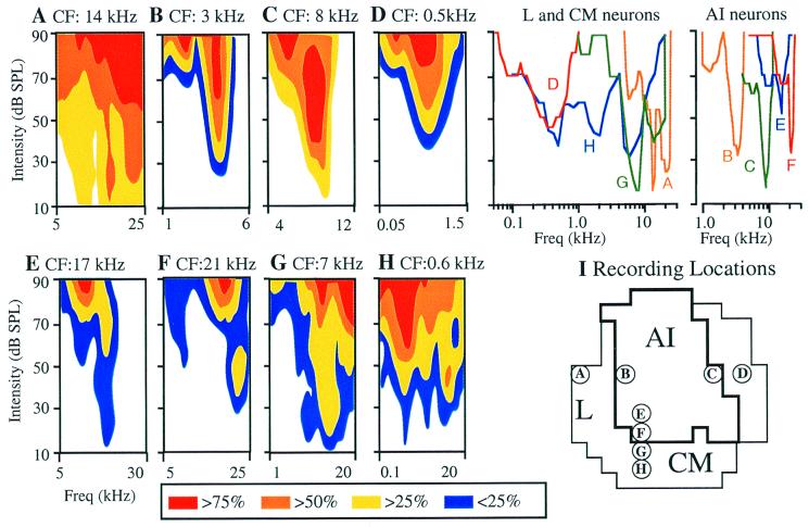
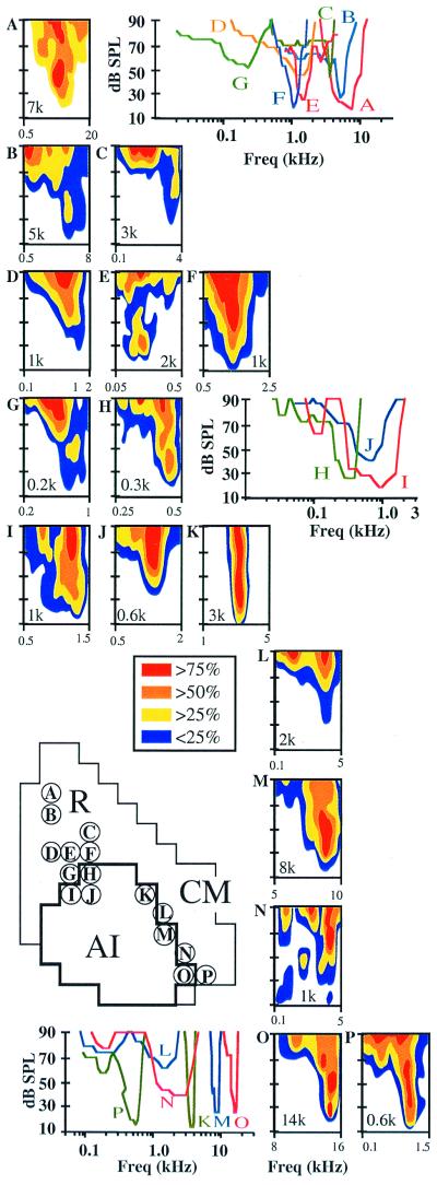
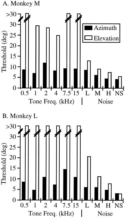
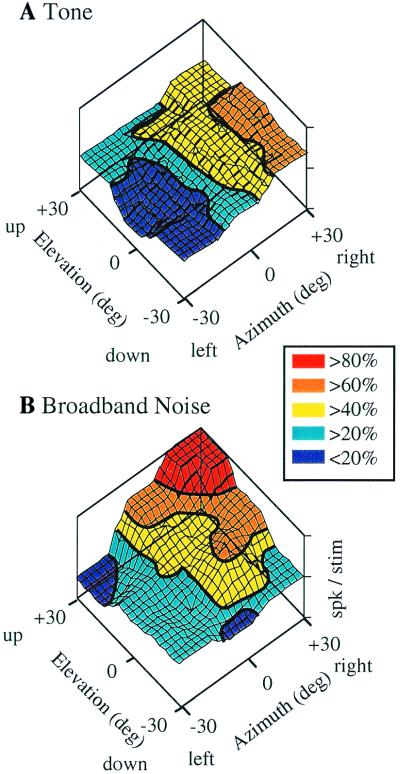
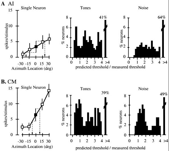
The patterns of cortico-cortical and cortico-thalamic connections of auditory cortical areas in the rhesus monkey have led to the hypothesis that acoustic information is processed in series and in parallel in the primate auditory cortex. Recent physiological experiments in the behaving monkey indicate that the response properties of neurons in different cortical areas are both functionally distinct from each other, which is indicative of parallel processing, and functionally similar to each other, which is indicative of serial processing. Thus, auditory cortical processing may be similar to the serial and parallel "what" and "where" processing by the primate visual cortex. If "where" information is serially processed in the primate auditory cortex, neurons in cortical areas along this pathway should have progressively better spatial tuning properties. This prediction is supported by recent experiments that have shown that neurons in the caudomedial field have better spatial tuning properties than neurons in the primary auditory cortex. Neurons in the caudomedial field are also better than primary auditory cortex neurons at predicting the sound localization ability across different stimulus frequencies and bandwidths in both azimuth and elevation. These data support the hypothesis that the primate auditory cortex processes acoustic information in a serial and parallel manner and suggest that this may be a general cortical mechanism for sensory perception.


Figures

Figure 1
Figure 1
Frequency response areas of single auditory cortical neurons. Responses were recorded to 50-ms tone stimuli (3-ms rise/fall) presented at 16 different intensity levels [10- to 90-dB sound pressure level (SPL)] at 31 different frequencies spanning 2–5 octaves from a free-field speaker located directly opposite to the contralateral ear. The color corresponds to the percent of the maximum response recorded in that neuron. White areas correspond to areas where the activity was not significantly greater than the spontaneous rate. Each FRA shows the response of a single neuron from the cortical location shown in I. The frequency range was customized for each neuron and therefore will vary between panels. The 25% contour (50% for the neuron shown in A) is reproduced on the same frequency axis to allow comparisons of the frequency bandwidth across neurons (Upper Right), and the CF is given above each FRA. (I) Dorsal view of the recording locations for each neuron. The heavy line shows the physiological boundaries of AI. Thin lines show the region investigated in the study. Circled letters correspond to the different panels shown in the figure. Note the differences in frequency tuning between neurons in AI and other cortical fields. Adapted from ref. .
Figure 2
Figure 2
FRAs recorded in the three different auditory cortical areas in a second monkey. (AG) FRAs from single neurons recorded in R. (HJ) FRAs recorded at the rostral border of AI. (K, M, and O) Neurons recorded at the medial border of AI. (L, N, and P) Neurons recorded in CM near the AI-CM border. The characteristic frequency is shown within each FRA. Other conventions are as in Fig. 1. Adapted from ref. .
Figure 3
Figure 3
Sound localization thresholds across stimulus frequencies and bandwidths. Thresholds are shown for localization in azimuth (solid bars) and elevation (open bars). Thresholds could not be defined if they were greater than 30° (broken lines). Noise stimuli consisted of 1-octave band-passed nose (L: 750–1,500 Hz; M: 3,000–6,000 Hz; H: 5,000–10,000 Hz) and broadband noise (NS). Adapted from ref. .
Figure 4
Figure 4
Spatial response profiles of an AI neuron. PSTHs are shown in their relative position from the monkey's perspective (rightward PSTHs correspond to stimuli presented to the right of midline). Numbers above the most eccentric PSTHs correspond to the location in degrees (azimuth, elevation). Each PSTH shows the responses over 10 trials. Tone (A) and noise (B) stimuli were presented on randomly interleaved trials. In the color-coded three-dimensional plots the response was normalized by the maximum response recorded for that neuron to any of the 17 locations using either the tone or noise stimulus. The magnitude of the response at each azimuth and elevation is shown by the height of the contour. Heavy lines show regions with the same activity (iso-response contours).
Figure 5
Figure 5
Three-dimensional reconstructions of spatial responses from a representative CM neuron. Conventions are as in Fig. 4. This neuron had a lower response to tone stimuli, and a more shallow response as a function of stimulus location (A). The response to noise stimuli (B) showed greater modulation as a function of stimulus location, and the slope of this response contour was not aligned with either the elevation or azimuth axis. This indicates that this neuron contained information for both the azimuth and elevation of the stimulus.
Figure 6
Figure 6
Predictions of behavioral performance by single neurons. The mean and standard deviation of the response of a single neuron as a function of the stimulus azimuth (0° elevation) are shown for an AI neuron (A) and a CM neuron (B). (Left) ▪ notes the response from the speaker located directly in front of the monkey. The ability of the neuron to predict the behavior was calculated as the distance in azimuth that corresponded to one standard deviation from the mean response at 0° (dashed lines). This prediction was tested against the behavior by dividing the predicted threshold by the measured threshold. (Center) The frequency distribution of this ratio when predicting thresholds in azimuth for tone stimuli measured across 353 AI neurons (A) and 118 CM neurons (B). Neurons that had a prediction greater than four times the measured threshold are shown in the right most bin. Ratios of 1.0 correspond to perfect predictions. (Right) The ratios when predicting thresholds in azimuth for noise stimuli. Adapted from ref. .
Figure 7
Figure 7
Mean and standard deviation for the predicted/measured ratio pooled across either all neurons measured in that cortical area (open bars) or restricted to only the neurons in that cortical area that had significant correlation between the neuronal activity and the spatial location for at least one stimulus (closed bars). Each bar represents the mean of the azimuth and elevation predictions for tone and noise stimuli (21 ratios total). Dashed line is through 1.0 (perfect prediction). Only the pooled spatially sensitive CM neurons had a ratio that was not significantly different from the behavior. Adapted from ref. .

References

    1. Middlebrooks J C, Green D M. Annu Rev Psychol. 1991;42:135–159. - PubMed
    1. Wightman F L, Kistler D J. J Acoust Soc Am. 1989;85:868–878. - PubMed
    1. Pralong D, Carlile S. J Acoust Soc Am. 1994;95:3435–3444. - PubMed
    1. Joris P X, Yin T C. J Neurophysiol. 1995;73:1043–1062. - PubMed
    1. Yin T C, Chan J C. J Neurophysiol. 1990;64:465–488. - PubMed

Publication types

LinkOut - more resources