. 2000 Nov;182(22):6358-65.
doi: 10.1128/JB.182.22.6358-6365.2000.

Detection of mRNA transcripts and active transcription in persistent Mycobacterium tuberculosis induced by exposure to rifampin or pyrazinamide

Affiliations

Detection of mRNA transcripts and active transcription in persistent Mycobacterium tuberculosis induced by exposure to rifampin or pyrazinamide

Y Hu et al. J Bacteriol. 2000 Nov.

Abstract

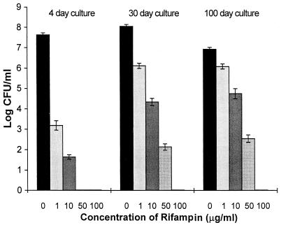
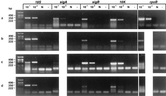
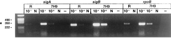
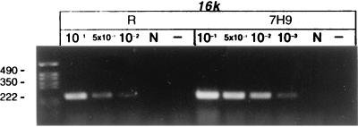
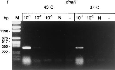
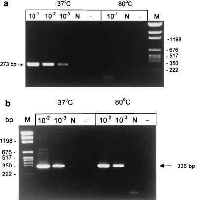
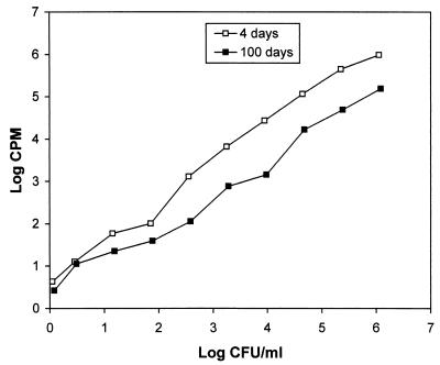
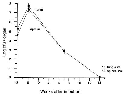
Mycobacterium tuberculosis can persist in an altered physiological state for many years after initial infection, and it may reactivate to cause active disease. An analogous persistent state, possibly consisting of several different subpopulations of bacteria, may arise during chemotherapy; this state is thought to be responsible for the prolonged period required for effective chemotherapy. Using two models of drug-induced persistence, we show that both microaerophilic stationary-phase M. tuberculosis treated with a high dose of rifampin in vitro and pyrazinamide-induced persistent bacteria in mice are nonculturable yet still contain 16S rRNA and mRNA transcripts. Also, the in vitro persistent, plate culture-negative bacteria incorporate radioactive uridine into their RNA in the presence of rifampin and can rapidly up-regulate gene transcription after the replacement of the drug with fresh medium and in response to heat shock. Our results show that persistent M. tuberculosis has transcriptional activity. This finding provides a molecular basis for the rational design of drugs targeted at persistent bacteria.


Figures

FIG. 1
FIG. 1
Effects of different concentrations of rifampin on the viability of log-phase and stationary-phase cultures of M. tuberculosis H37Rv. Log-phase (4-day) and stationary-phase (30- and 100-day) cultures were incubated with 1, 10, 50, and 100 μg of rifampin ml−1 for 5 days. After washing of the cultures, viability was estimated by examination of CFU counts. Values shown are averages of three independent experiments.
FIG. 2
FIG. 2
Detection of mRNA in M. tuberculosis H37Rv by RT-PCR before and after treatment with antituberculosis agents in vitro and in vivo. (a) RNA was extracted from 100-day cultures before rifampin treatment (106 CFU per RT reaction). (b) After rifampin (100 μg/ml) treatment for 5 days (0 CFU; same volume of culture used for RT as in panel a). (c) RNA was extracted from infected mice before antituberculosis drug treatment; week 0 (106 CFU per RT). (d) After antituberculosis drug treatment in mice; week 14 (0 CFU; same volume of tissue used for RT as in panel c). (e) The 100-day cultures were treated with rifampin (100 μg/ml) for 5 days. The cells were washed three times to remove rifampin and then incubated with 7H9 broth for 12 h. RT-PCR was performed with sigA, sigB, rpoB, and 16K primers before (R) and after (7H9) resuscitation with 7H9 medium. (f) The 100-day cultures were treated with rifampin for 5 days, washed free of rifampin, and immediately heat shocked at 45°C for 30 min compared to incubation at 37°C for 30 min. Detected genes are shown above lanes; the PCR product sizes are as follows: sigA, 306 bp; sigB, 273 bp; rpoB, 307 bp; dnaK, 275 bp; 16K (hspX), 242 bp; 16S, 336 bp. 10−1, 5 × 10⁻¹, 10−2, and 10−3 indicate that 10-, 50-, 100-, and 1,000-fold-diluted cDNAs were used for PCR. N, DNase I-treated RNA but no RT enzyme. −, PCR control with each primer and 10 μl of water instead of template. The following limits of detection (gene copies) were estimated by DNA PCR: 16S, 105 (equivalent to 100 CFU of a log-phase growth culture); sigA, 107; sigB, 106; 16K, 106; rpoB, 106; dnaK, 106 (equivalent to ∼104 CFU).
FIG. 2
FIG. 2
Detection of mRNA in M. tuberculosis H37Rv by RT-PCR before and after treatment with antituberculosis agents in vitro and in vivo. (a) RNA was extracted from 100-day cultures before rifampin treatment (106 CFU per RT reaction). (b) After rifampin (100 μg/ml) treatment for 5 days (0 CFU; same volume of culture used for RT as in panel a). (c) RNA was extracted from infected mice before antituberculosis drug treatment; week 0 (106 CFU per RT). (d) After antituberculosis drug treatment in mice; week 14 (0 CFU; same volume of tissue used for RT as in panel c). (e) The 100-day cultures were treated with rifampin (100 μg/ml) for 5 days. The cells were washed three times to remove rifampin and then incubated with 7H9 broth for 12 h. RT-PCR was performed with sigA, sigB, rpoB, and 16K primers before (R) and after (7H9) resuscitation with 7H9 medium. (f) The 100-day cultures were treated with rifampin for 5 days, washed free of rifampin, and immediately heat shocked at 45°C for 30 min compared to incubation at 37°C for 30 min. Detected genes are shown above lanes; the PCR product sizes are as follows: sigA, 306 bp; sigB, 273 bp; rpoB, 307 bp; dnaK, 275 bp; 16K (hspX), 242 bp; 16S, 336 bp. 10−1, 5 × 10⁻¹, 10−2, and 10−3 indicate that 10-, 50-, 100-, and 1,000-fold-diluted cDNAs were used for PCR. N, DNase I-treated RNA but no RT enzyme. −, PCR control with each primer and 10 μl of water instead of template. The following limits of detection (gene copies) were estimated by DNA PCR: 16S, 105 (equivalent to 100 CFU of a log-phase growth culture); sigA, 107; sigB, 106; 16K, 106; rpoB, 106; dnaK, 106 (equivalent to ∼104 CFU).
FIG. 3
FIG. 3
mRNA as a correlate of viability, analyzed by limiting dilution RT-PCR of heat-killed and viable 100-day cultures of M. tuberculosis. (a) sigB mRNA; (b) 16S rRNA. Lanes N and −, as in Fig. 2; lane M, molecular weight markers.
FIG. 4
FIG. 4
Kinetic analysis of [3H]uridine incorporation by log-phase and stationary-phase M. tuberculosis. Log-phase (4-day) and stationary-phase (100-day) cultures were serially diluted fivefold. [3H]uridine was added to the cultures, and RNA was extracted after 20 h of incubation. The results were confirmed in three independent experiments.
FIG. 5
FIG. 5
Viability of M. tuberculosis H37Rv in vivo before and after treatment with antituberculosis agents in the mouse Cornell model. The results of a single experiment are shown, with viability expressed as log CFU counts per organ (spleen or lung). Mice were infected intravenously at week −2, and the infection allowed to progress for 2 weeks prior to treatment with pyrazinamide and isoniazid for 14 weeks (weeks 0 to 14). At week 14, 2 mice yielded a positive organ culture, one for lung only (1/8 lung +ve) and one for spleen only (1/8 spleen +ve). The remaining 6 mice at week 14 + 1 were all culture negative, and their spleens and lungs were frozen in one-fifth portions for RNA extraction and RT-PCR analysis. Not shown is the bacteriological relapse in 21 of 23 mice after steroid treatment given at week 14 + 8, indicating restoration of culturability and hence rescue from dormancy.

References

    1. Bloom B R, McKinney J D. The death and resurrection of tuberculosis. Nat Med. 1999;5:872–874. - PubMed
    1. Butcher P D, Mangan J A, Monahan I M. Intracellular gene expression: analysis of RNA from mycobacteria in macrophages using RT-PCR. Methods Mol Biol. 1998;101:285–306. - PubMed
    1. Cole S T, Brosch R, Parkhill J, Garnier T, Churcher C, Harris D, Gordon S V, Eiglmeier K, Gas S, Barry III C E, Tekaia F, Badcock K, Basham D, Brown D, Chillingworth T, Connor R, Davies R, Devlin K, Feltwell T, Gentles S, Hamlin N, Holroyd S, Hornsby T, Jagels K, Krogh A, McLean J, Moule S, Murphy L, Oliver K, Osborne J, Rajandream M-A, Rogers J, Rutter S, Seeger K, Skelton J, Squares R, Squares S, Sulston J E, Taylor K, Whitehead S, Barrell B G. Deciphering the biology of Mycobacterium tuberculosis from the complete genome sequence. Nature. 1998;393:537–544. - PubMed
    1. Cunningham A F, Spreadbury C L. Mycobacterial stationary phase induced by low oxygen tension: cell wall thickening and localization of the 16-kilodalton α-crystallin homolog. J Bacteriol. 1998;180:801–808. - PMC - PubMed
    1. De Wit D, Wootton M, Dhillon J, Mitchison D A. The bacterial DNA content of mouse organs in the Cornell model of dormant tuberculosis. Tuber Lung Dis. 1995;76:555–562. - PubMed

Publication types

MeSH terms