. 2000 Nov;182(22):6463-71.
doi: 10.1128/JB.182.22.6463-6471.2000.

Bacillus subtilis SMC is required for proper arrangement of the chromosome and for efficient segregation of replication termini but not for bipolar movement of newly duplicated origin regions

Affiliations

Bacillus subtilis SMC is required for proper arrangement of the chromosome and for efficient segregation of replication termini but not for bipolar movement of newly duplicated origin regions

P L Graumann. J Bacteriol. 2000 Nov.

Abstract

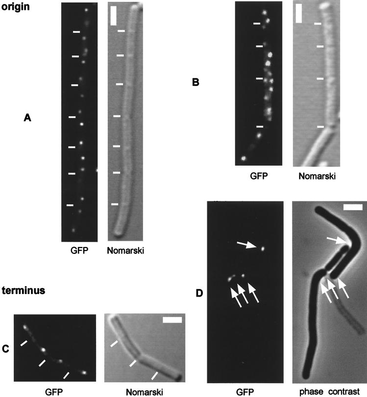
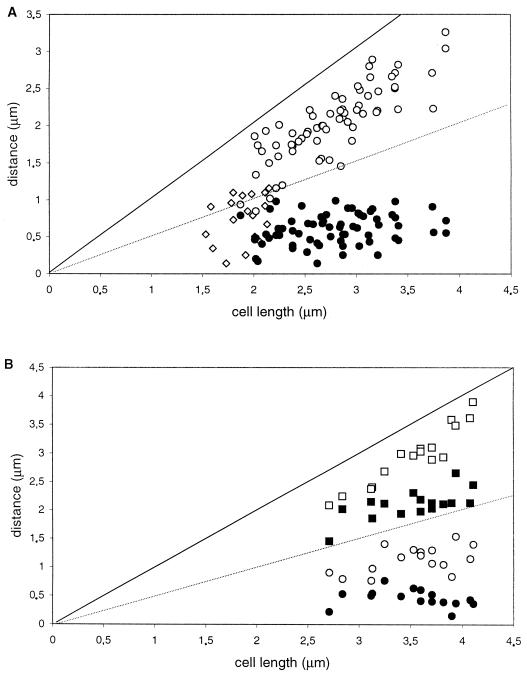
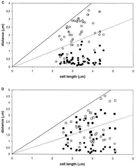
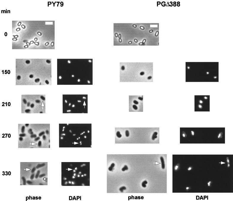
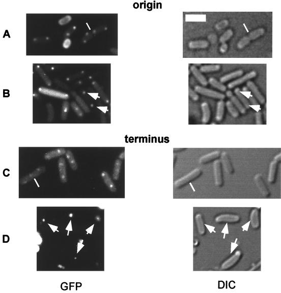
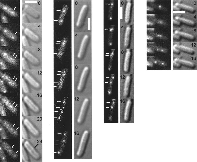
SMC protein is required for chromosome condensation and for the faithful segregation of daughter chromosomes in Bacillus subtilis. The visualization of specific sites on the chromosome showed that newly duplicated origin regions in growing cells of an smc mutant were able to segregate from each other but that the location of origin regions was frequently aberrant. In contrast, the segregation of replication termini was impaired in smc mutant cells. This analysis was extended to germinating spores of an smc mutant. The results showed that during germination, newly duplicated origins, but not termini, were able to separate from each other in the absence of SMC. Also, DAPI (4',6'-diamidino-2-phenylindole) staining revealed that chromosomes in germinating spores were able to undergo partial or complete replication but that the daughter chromosomes were blocked at a late stage in the segregation process. These findings were confirmed by time-lapse microscopy, which showed that after duplication in growing cells the origin regions underwent rapid movement toward opposite poles of the cell in the absence of SMC. This indicates that SMC is not a required component of the mitotic motor that initially drives origins apart after their duplication. It is also concluded that SMC is needed to maintain the proper layout of the chromosome in the cell and that it functions in the cell cycle after origin separation but prior to complete segregation or replication of daughter chromosomes. It is proposed here that chromosome segregation takes place in at least two steps: an SMC-independent step in which origins move apart and a subsequent SMC-dependent step in which newly duplicated chromosomes condense and are thereby drawn apart.


Figures

FIG. 1
FIG. 1
Fluorescence microscopy of B. subtilis cells producing GFP-LacI and carrying tandem copies of lacO near the origin (A and B) or terminus (C and D) of replication. (A) AT63 (wild type). (B) PG63 (smc::kan). (C) AT62 (wild type). (D) PG6 (smc::kan). The white lines indicate septa, which were visualized by differential interference contrast (Nomarski) microscopy. The scale bar (thick white line) represents 2 μm. Cells were grown in rich medium at 23°C, and images were collected during the mid-exponential phase of growth.
FIG. 2
FIG. 2
Distance of fluorescent foci from the cell pole as a function of cell size. (A and B) AT63 (GFP-LacI, lacO cassette at 359°). (C and D) PG63 (smc::kan, GFP-LacI, lacO cassette at 359°). Cells were grown in rich medium at 23°C, and images were obtained during the mid-exponential phase of growth. Panels A and C show cells with one (⧫) or two (● and ○) fluorescent foci; panels B and D show cells with four foci (●, ○, ■, and □, with “●” being the closest and “□” being the farthest focus from the pole chosen for measurements). The measurements in panels C and D do not include very large cells, in which origin signals tended to be even more random than in small cells.
FIG. 2
FIG. 2
Distance of fluorescent foci from the cell pole as a function of cell size. (A and B) AT63 (GFP-LacI, lacO cassette at 359°). (C and D) PG63 (smc::kan, GFP-LacI, lacO cassette at 359°). Cells were grown in rich medium at 23°C, and images were obtained during the mid-exponential phase of growth. Panels A and C show cells with one (⧫) or two (● and ○) fluorescent foci; panels B and D show cells with four foci (●, ○, ■, and □, with “●” being the closest and “□” being the farthest focus from the pole chosen for measurements). The measurements in panels C and D do not include very large cells, in which origin signals tended to be even more random than in small cells.
FIG. 3
FIG. 3
Time course of germinating spores from strain PY79 (wild type) and PGΔ388 (smc::kan). Samples of cells at the indicated times after the start of germination were fixed and stained with DAPI prior to image acquisition. DAPI images of purified spores are not shown since spores show a strong blue autofluorescence. The scale bars (thick white lines) represent 2 μm. The arrows indicate partially (210-min time point) and completely (270- and 330-min time points) separated nucleoids.
FIG. 4
FIG. 4
Fluorescence and Nomarski DIC images of germinating spores of cells producing GFP-LacI and carrying tandem copies of lacO. Images were collected 1.5 h after resuspension in salt buffer. Panels A and C show wild-type cells (AT63 and AT62, respectively) and panels B and D show the smc mutant cells (PG63 and PG6, respectively) containing the lacO cassette near the origin (359°; A and B) or near the terminus (180°; C and D). The scale bars (thick white lines) represent 2 μm, the arrows indicate the position of fluorescent foci, and the white bars show the position of the septa.
FIG. 5
FIG. 5
Time-lapse microscopy. Cells from the smc mutant PG63, which produces GFP-LacI and contains the lacO cassette at 359°, were grown on agarose pads containing S750 medium at room temperature. The left-hand columns show fluorescence from GFP, and the right-hand columns show Nomarski DIC images. The numbers indicate the time in minutes at which the images were acquired. The scale bar (thick white line) represents 2 μm. The white lines indicate the position of fluorescent foci; the black lines indicate the emergence of a septum.

Similar articles

Cited by

References

    1. Akhmedov A T, Frei C, Tsai-Pflugfelder M, Kemper B, Gasser S M, Jessberger R. Structural maintenance of chromosomes protein C-terminal domains bind preferentially to DNA with secondary structure. J Biol Chem. 1998;273:24088–24094. - PubMed
    1. Blat Y, Kleckner N. Cohesins bind to preferential sites along yeast chromosome III, with differential regulation along arms versus the centric region. Cell. 1999;98:249–259. - PubMed
    1. Britton R A, Lin D C-H, Grossman A D. Characterization of a procaryotic SMC protein involved in chromosome partitioning. Genes Dev. 1998;12:1254–1259. - PMC - PubMed
    1. Callister H, Wake R G. Completion of the replication and division cycle in temperature-sensitive DNA initiation mutants of Bacillus subtilis 168 at the non-permissive temperature. J Mol Biol. 1977;117:71–84. - PubMed
    1. Glaser P, Sharpe M E, Raether B, Perego M, Ohlsen K, Errington J. Dynamic, mitotic-like behavior of a bacterial protein required for accurate chromosome partitioning. Genes Dev. 1997;11:1160–1168. - PubMed

Publication types

LinkOut - more resources