Comparative Study
. 2000 Nov;182(22):6482-9.
doi: 10.1128/JB.182.22.6482-6489.2000.

Development and dynamics of Pseudomonas sp. biofilms

Affiliations
Comparative Study

Development and dynamics of Pseudomonas sp. biofilms

T Tolker-Nielsen et al. J Bacteriol. 2000 Nov.

Abstract

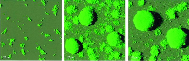
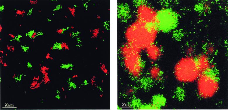
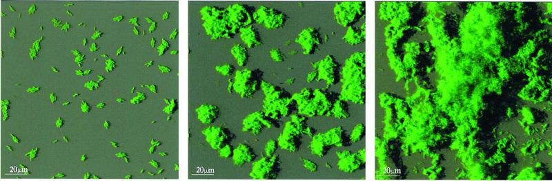
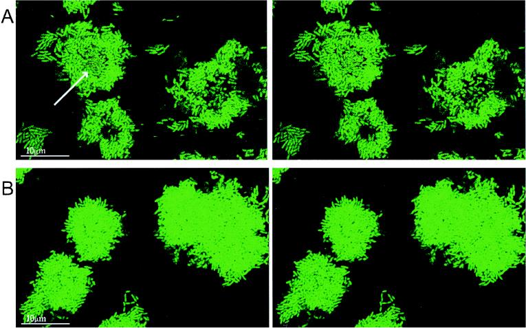
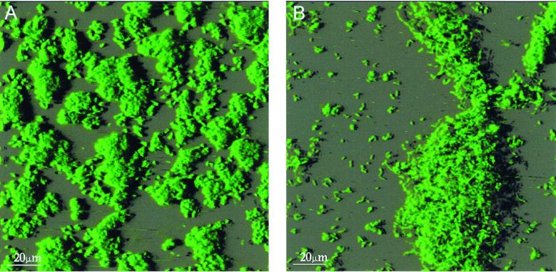
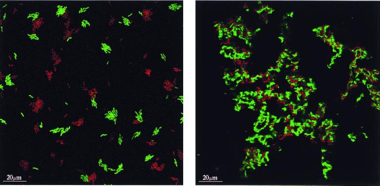
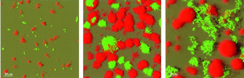
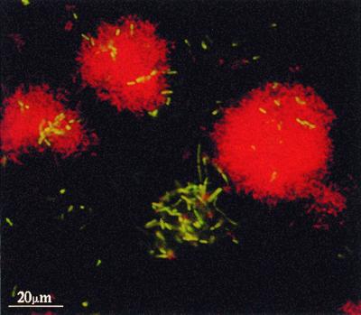
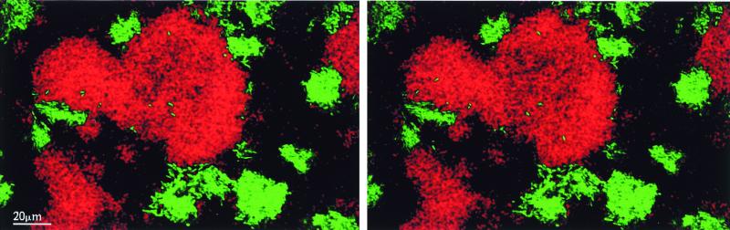
Pseudomonas sp. strain B13 and Pseudomonas putida OUS82 were genetically tagged with the green fluorescent protein and the Discosoma sp. red fluorescent protein, and the development and dynamics occurring in flow chamber-grown two-colored monospecies or mixed-species biofilms were investigated by the use of confocal scanning laser microscopy. Separate red or green fluorescent microcolonies were formed initially, suggesting that the initial small microcolonies were formed simply by growth of substratum attached cells and not by cell aggregation. Red fluorescent microcolonies containing a few green fluorescent cells and green fluorescent microcolonies containing a few red fluorescent cells were frequently observed in both monospecies and two-species biofilms, suggesting that the bacteria moved between the microcolonies. Rapid movement of P. putida OUS82 bacteria inside microcolonies was observed before a transition from compact microcolonies to loose irregularly shaped protruding structures occurred. Experiments involving a nonflagellated P. putida OUS82 mutant suggested that the movements between and inside microcolonies were flagellum driven. The results are discussed in relation to the prevailing hypothesis that biofilm bacteria are in a physiological state different from planktonic bacteria.


Figures

FIG. 1
FIG. 1
The spatial structures in a developing B13(GF) biofilm were studied by the use of CSLM. Shadow projection micrographs recorded in a 1 (left)-, 3 (middle)-, and 5 (right)-day-old biofilm are shown.
FIG. 2
FIG. 2
The location of green and red fluorescent bacteria within horizontal sections of a developing B13(GF)-B13(RF) biofilm was studied by the use of CSLM. Optical section micrographs recorded in a 1 (left)- and 5 (right)-day-old biofilm are shown.
FIG. 3
FIG. 3
The spatial structures in a developing OUS82(GF) biofilm were studied by the use of CSLM. Shadow projection micrographs recorded in a 1 (left)-, 3 (middle)-, and 5 (right)-day-old biofilm are shown.
FIG. 4
FIG. 4
The location of bacteria within horizontal sections of a 3-day-old OUS82(GF) biofilm (A) and a 3-day-old OUS82(Fla) biofilm (B) was studied by the use of CSLM. The left optical section micrographs were recorded 20 s before the right optical section micrographs. The mowing cells inside one of the OUS82(GF) microcolonies, which do not appear in the same position on the left and right micrographs, are pointed out.
FIG. 5
FIG. 5
The spatial structures in OUS82(GF) and OUS82(Fla) biofilms were studied by the use of CSLM. Shadow projection micrographs of a 7-day-old OUS82(Fla) biofilm (A) and a 7-day-old OUS82(GF) biofilm (B) are shown.
FIG. 6
FIG. 6
The location of green and red fluorescent bacteria within horizontal sections of a developing OUS82(GF)-OUS82(RF) biofilm was studied by the use of CSLM. Optical section micrographs recorded in a 1 (left)- and 5 (right)-day-old biofilm are shown.
FIG. 7
FIG. 7
The spatial structures in a developing B13(RF)-OUS82(GF) biofilm were studied by the use of CSLM. Shadow projection micrographs of a 1 (left)-, 2 (middle)-, and 5 (right)-day-old biofilm are shown.
FIG. 8
FIG. 8
The location of green and red fluorescent bacteria within horizontal sections of a developing B13(RF)-OUS82(GF) biofilm was studied by the use of CSLM. An optical section micrograph recorded in a 5-day-old biofilm is shown.
FIG. 9
FIG. 9
Movement of green fluorescent OUS82(GF) bacteria inside red fluorescent B13(RF) microcolonies was studied by the use of CSLM. The left optical section micrograph was recorded 1 min before the right optical section micrograph. The appearance of OUS82(GF) bacteria in different positions inside the B13(RF) microcolony on the two micrographs suggests that the OUS82(GF) bacteria moved inside the B13(RF) microcolony.

References

    1. Andersen J B, Sternberg C, Poulsen L K, Bjorn S P, Givskov M, Molin S. New unstable variants of green fluorescent protein for studies of transient gene expression in bacteria. Appl Environ Microbiol. 1998;64:2240–2246. - PMC - PubMed
    1. Bertani G. Studies on lysogenesis. I. The mode of phage liberation by lysogenic Escherichia coli. J Bacteriol. 1951;62:293–300. - PMC - PubMed
    1. Clark J D, Maaløe O. DNA replication and the cell cycle in Escherichia coli cells. J Mol Biol. 1967;23:99–112.
    1. Costerton J W. Introduction to biofilm. Int J Antimicrob Agents. 1999;11:217–221. - PubMed
    1. Costerton J W, Stewart P S, Greenberg E P. Bacterial biofilms: a common cause of persistent infections. Science. 1999;284:1318–1322. - PubMed

Publication types

MeSH terms

Associated data

LinkOut - more resources