. 2001 Feb 9;276(6):4063-9.
doi: 10.1074/jbc.M007419200. Epub 2000 Oct 27.

Munc18c regulates insulin-stimulated glut4 translocation to the transverse tubules in skeletal muscle

Affiliations

Munc18c regulates insulin-stimulated glut4 translocation to the transverse tubules in skeletal muscle

A H Khan et al. J Biol Chem. .

Abstract

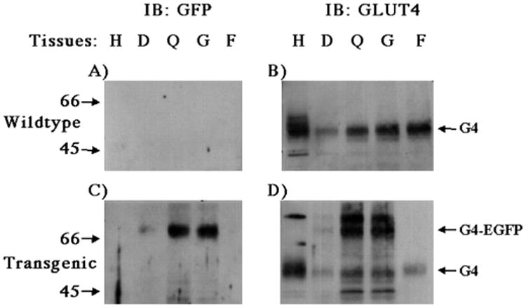
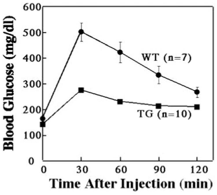
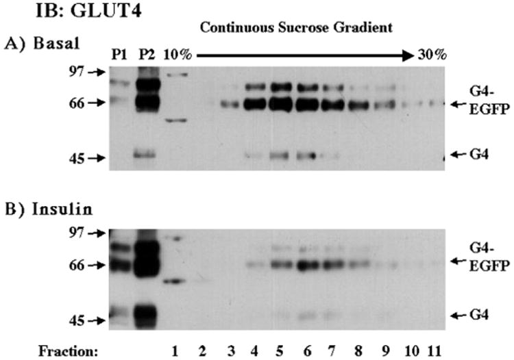
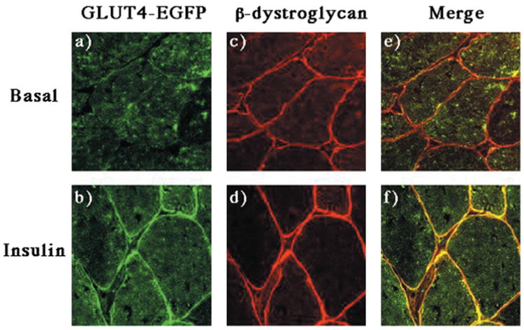
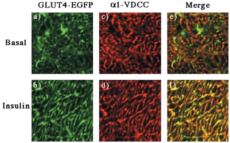
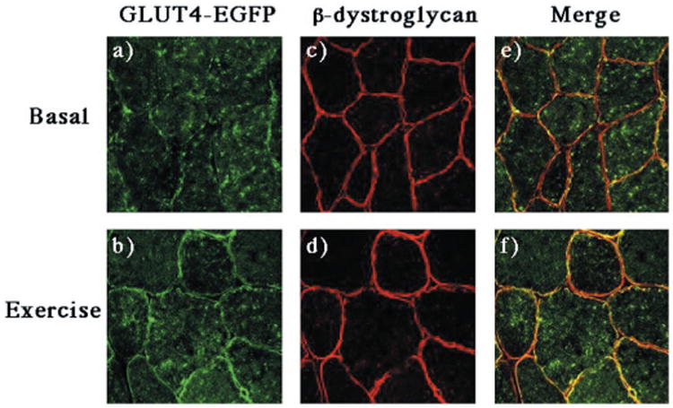
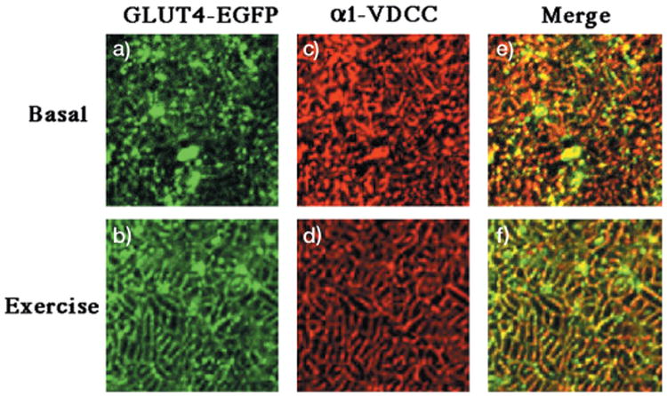
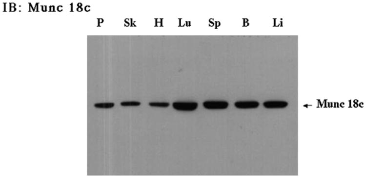
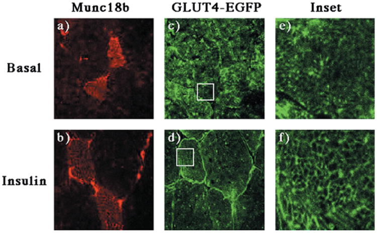
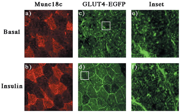
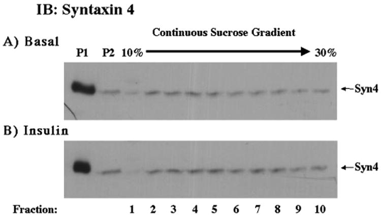
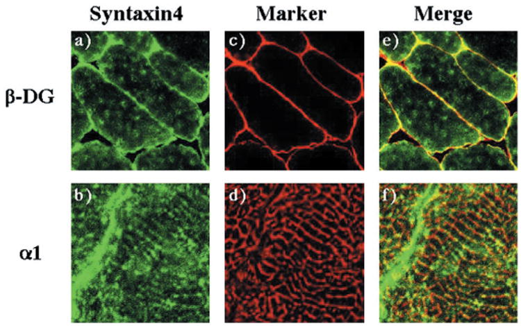
To examine the intracellular trafficking and translocation of GLUT4 in skeletal muscle, we have generated transgenic mouse lines that specifically express a GLUT4-EGFP (enhanced green fluorescent protein) fusion protein under the control of the human skeletal muscle actin promoter. These transgenic mice displayed EGFP fluorescence restricted to skeletal muscle and increased glucose tolerance characteristic of enhanced insulin sensitivity. The GLUT4-EGFP protein localized to the same intracellular compartment as the endogenous GLUT4 protein and underwent insulin- and exercise-stimulated translocation to both the sarcolemma and transverse-tubule membranes. Consistent with previous studies in adipocytes, overexpression of the syntaxin 4-binding Munc18c isoform, but not the related Munc18b isoform, in vivo specifically inhibited insulin-stimulated GLUT4-EGFP translocation. Surprisingly, however, Munc18c inhibited GLUT4 translocation to the transverse-tubule membrane without affecting translocation to the sarcolemma membrane. The ability of Munc18c to block GLUT4-EGFP translocation to the transverse-tubule membrane but not the sarcolemma membrane was consistent with substantially reduced levels of syntaxin 4 in the transverse-tubule membrane. Together, these data demonstrate that Munc18c specifically functions in the compartmentalized translocation of GLUT4 to the transverse-tubules in skeletal muscle. In addition, these results underscore the utility of this transgenic model to directly visualize GLUT4 translocation in skeletal muscle.


Figures

Fig. 1
Fig. 1. The human skeletal actin muscle promoter drives the tissue-specific expression of the GLUT4-EGFP fusion protein in transgenic mice
Tissues from wild-type (A and B) and GLUT4-EGFP trangenic (C and D) mice were isolated and subjected to immunoblotting as described under “Experimental Procedures.” Tissue extracts (5 μg) were prepared from the heart (H), diaphragm (D), quadriceps (Q), gastrocnemius (G), and epididymal adipose depot (F) and subjected to SDS-polyacrylamide gel electrophoresis. The samples were then immunoblotted with the GFP antibody (A and C) or the GLUT4 antibody (B and D). These are representative immunoblots performed 6 times from four different independent transgenic mice.
Fig. 2
Fig. 2. The skeletal muscle specific GLUT4-EGFP transgenic mice display greater insulin sensitivity than wild-type mice
Wild-type and GLUT4-EGFP transgenic mice were fasted overnight and then given an intraperitoneal injection of glucose (2 g/kg). Plasma glucose levels were then determined at the times indicated as described under “Experimental Procedures.” The average and S.E. of the mean are shown for 7 wild-type (WT) and 10 GLUT4-EGFP (TG) transgenic mice.
Fig. 3
Fig. 3. Insulin stimulation results in the skeletal muscle surface membrane translocation of both the endogenous GLUT4 and GLUT4-EGFP fusion proteins
GLUT4-EGFP transgenic mice were fasted overnight and either left untreated (A) or stimulated with insulin (B) as described under “Experimental Procedures.” The hind-quarter skeletal muscle was dissected and subjected to differential and sucrose velocity centrifugation as described under “Experimental Procedures.” The surface membrane fractions (P1 and P2) as well as the intracellular membrane fractions (fractions 1–11) were immunoblotted with the GLUT4 antibody. These are representative immunoblots performed from four different independent pairs of transgenic mice.
Fig. 4
Fig. 4. Insulin stimulation results in the translocation of GLUT4-EGFP to the sarcolemma membrane
GLUT4-EGFP transgenic mice were fasted overnight and either left untreated (panels a, c, and e) or stimulated with insulin (panels b, d, and f) as described under “Experimental Procedures.” The quadriceps muscle was frozen, sliced, and incubated with the β-dystroglycan monoclonal antibody and secondary donkey anti-mouse IgG conjugated to Texas Red (panels c and d). Sections were visualized by confocal fluorescent microscopy (×63). These are representative fields from three independent experiments.
Fig. 5
Fig. 5. Insulin stimulation results in the translocation of GLUT4-EGFP to the transverse-tubule membranes
GLUT4-EGFP transgenic mice were fasted overnight and either left untreated (panels a, c, and e) or stimulated with insulin (panels b, d, and f) as described under “Experimental Procedures.” The quadriceps muscle was frozen, sliced, and incubated with the monoclonal antibody directed against the α1 subunit of the α1-VDCC using the Monoclonal on Mouse kit according to the manufacturers instructions (panels c and d). Sections were visualized by confocal fluorescent microscopy (×63) and then further magnified 11 times. The width of each panel is ∼13 μm. These are representative fields from three independent experiments.
Fig. 6
Fig. 6. Exercise results in the translocation of GLUT4-EGFP to the sarcolemma membrane
GLUT4-EGFP transgenic mice were fasted overnight and either left untreated (panels a, c, and e) or run for 60 min on a treadmill (panels b, d, and f) as described under “Experimental Procedures.” The quadriceps muscle was frozen, sliced, and incubated with the β-dystroglycan monoclonal antibody and secondary donkey anti-mouse IgG conjugated to Texas Red (panels c and d). Sections were visualized by confocal fluorescent microscopy (×63). These are representative fields from two independent experiments.
Fig. 7
Fig. 7. Exercise stimulation results in the translocation of GLUT4-EGFP to the transverse-tubule membranes
GLUT4-EGFP transgenic mice were fasted overnight and either left untreated (panels a, c, and e) or run for 60 min on a treadmill (panels b, d, and f) as described under “Experimental Procedures.” The quadriceps muscle was frozen, sliced, and incubated with the monoclonal antibody directed against the α1 subunit of the α1-VDCC using the Monoclonal on Mouse kit according to manufacturers instructions (panels c and d). Sections were visualized by confocal fluorescent microscopy (×63) and then further magnified 11 times. The width of each panel is ∼μm. These are representative fields from three independent experiments.
Fig. 8
Fig. 8. Munc18c is expressed in a number of different tissues, including skeletal muscle
Tissues from a wild-type C57Bl6 mouse were isolated and subjected to immunoblotting as described under “Experimental Procedures.” Tissue extracts (40 μg) were prepared from the following tissues: pancreas (P), skeletal muscle (Sk), heart (H), lung (Lu), spleen (Sp), brain (B), and liver (Li) and subjected to SDS-polyacrylamide gel electrophoresis. The samples were immunoblotted with the Munc18c rabbit polyclonal antibody.
Fig. 9
Fig. 9. Expression of Munc18b has no effect on the insulin stimulated translocation of GLUT4-EGFP to either the sarcolemma or transverse-tubule membranes
The quadricep muscle of GLUT4-EGFP transgenic mice was injected with a recombinant adenovirus encoding for the Munc18b protein as described under “Experimental Procedures.” Four days following injection, the mice were fasted overnight and either left untreated (panels a, c, and e) or stimulated with insulin (panels b, d, and f). The quadriceps muscle was frozen, sliced, and incubated with the Munc18b antibody and secondary donkey anti-rabbit IgG conjugated to Texas Red (panels a and b). Sections were visualized by confocal fluorescent microscopy (×63) and then further magnified 11 times (insets). The width of the box is ∼μm. These are representative fields from three independent experiments.
Fig. 10
Fig. 10. Expression of Munc18c has no effect on the insulin stimulated translocation of GLUT4-EGFP to the sarcolemma membrane but inhibits GLUT4-EGFP translocation to the transverse-tubules
The quadricep muscle of GLUT4-EGFP transgenic mice was injected with a recombinant adenovirus encoding for the Munc18c protein as described under “Experimental Procedures.” Four days following injection, the mice were fasted overnight and either left untreated (panels a, c, and e) or stimulated with insulin (panels b, d, and f). The quadriceps muscle was frozen, sliced, and incubated with the Munc18c antibody and secondary donkey anti-rabbit IgG conjugated to Texas Red (panels a and b). Sections were visualized by confocal fluorescent microscopy (×63) and then further magnified 11 times (insets). The width of the box is ∼μm. These are representative fields from four independent experiments.
Fig. 11
Fig. 11. Syntaxin 4 localizes primarily to the P1 fraction, and to a lesser extent to the P2 and sucrose gradient fractions, and does not translocate in response to insulin
Mice were treated as described under “Experimental Procedures.” 100 μg of the P1 and P2 fractions and 100 μl of each sucrose gradient fraction were subjected to SDS-polyacrylamide gel electrophoresis. The samples were then immunoblotted with a digoxigenin-labeled sheep Syntaxin 4 antibody and anti-digoxigenin POD Fab fragments (Roche Molecular Biochemicals, Germany).
Fig. 12
Fig. 12. Syntaxin 4 is localized to both the sarcolemma and transverse-tubule membranes in skeletal muscle
The quadriceps muscle from wild-type mice was frozen, sliced, and labeled with the sheep syntaxin 4 antibody (panels a and b), the β-dystroglycan monoclonal antibody (panel c), or the monoclonal α1-VDCC antibody (panel d) as described under “Experiment Procedures.” Sections were visualized by confocal fluorescent microscopy (×63). The transverse-tubules immunofluorescence was magnified 11 times from the confocal image. The width of each panel is ∼μm. These are representative fields from two independent experiments.

Similar articles

Cited by

References

    1. Kandror KV, Pilch PF. Am J Physiol. 1996;271:E1–E14. - PubMed
    1. Czech MP, Corvera S. J Biol Chem. 1999;274:1865–1868. - PubMed
    1. Goodyear LJ, Kahn BB. Annu Rev Med. 1998;49:235–261. - PubMed
    1. Birnbaum MJ, James DE. Curr Opin Endocrinol. 1995;2:383–391.
    1. Cheatham B, Vlahos CJ, Cheatham L, Wang L, Blenis J, Kahn CR. Mol Cell Biol. 1994;14:4902–4911. - PMC - PubMed

Publication types