. 2000 Nov;66(11):4655-61.
doi: 10.1128/AEM.66.11.4655-4661.2000.

Isolation and use of a homologous histone H4 promoter and a ribosomal DNA region in a transformation vector for the oil-producing fungus Mortierella alpina

Affiliations

Isolation and use of a homologous histone H4 promoter and a ribosomal DNA region in a transformation vector for the oil-producing fungus Mortierella alpina

D A Mackenzie et al. Appl Environ Microbiol. 2000 Nov.

Abstract

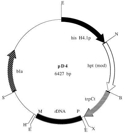
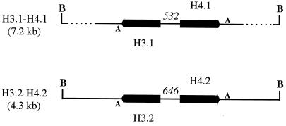
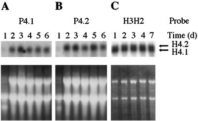
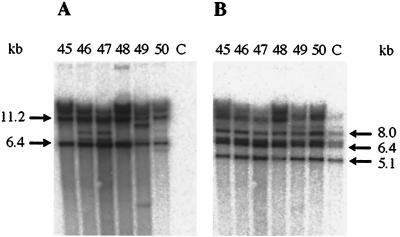
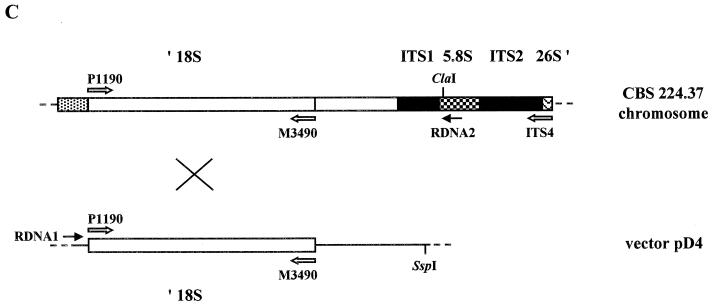
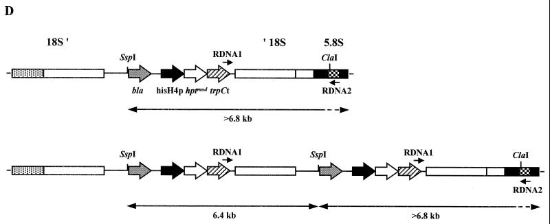
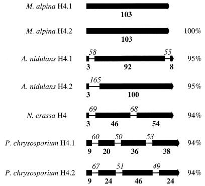
Mortierella alpina was transformed successfully to hygromycin B resistance by using a homologous histone H4 promoter to drive gene expression and a homologous ribosomal DNA region to promote chromosomal integration. This is the first description of transformation in this commercially important oleaginous organism. Two pairs of histone H3 and H4 genes were isolated from this fungus. Each pair consisted of one histone H3 gene and one histone H4 gene, transcribed divergently from an intergenic promoter region. The pairs of encoded histone H3 or H4 proteins were identical in amino acid sequence. At the DNA level, each histone H3 or H4 open reading frame showed 97 to 99% identity to its counterpart but the noncoding regions had little sequence identity. Unlike the histone genes from other filamentous fungi, all four M. alpina genes lacked introns. During normal vegetative growth, transcripts from the two histone H4 genes were produced at approximately the same level, indicating that either histone H4 promoter could be used in transformation vectors. The generation of stable, hygromycin B-resistant transformants required the incorporation of a homologous ribosomal DNA region into the transformation vector to promote chromosomal integration.


Figures

FIG. 1
FIG. 1
Map of the M. alpina transformation vector pD4. The 1-kb EcoRI-NcoI M. alpina histone H4.1 promoter fragment from strain CBS 528.72 also contains the histone H3.1 promoter and ORF. The hygromycin B resistance gene is the modified version (hptmod) which lacks the internal EcoRI and NcoI sites that are present in the wild-type gene of pAN7-1 (33). The 700-bp BamHI-XbaI fragment contains the A. nidulans trpC transcription terminator region (trpCt). The positions of the two rDNA primers P1190 and M3490, which were used to generate the M. alpina 18S rDNA fragment, are indicated as P and M, respectively. bla, ampicillin resistance gene. Restriction sites: B, BamHI; E, EcoRI; H, HindIII; N, NcoI; S, SspI; X, XbaI.
FIG. 2
FIG. 2
Organization of the two pairs of histone H3–H4 genes in M. alpina CBS 528.72. Heavy arrows indicate the position and direction of each ORF. The sizes of the promoter regions in nucleotides are given in italics. A, the consensus poly(A) addition signal AATAAA; B, BamHI site.
FIG. 3
FIG. 3
Transcription of the two histone H4 genes from M. alpina CBS 528.72. (A and B) Total RNA (20 μg per lane), which had been isolated from PDB-grown cultures harvested on the days indicated, was probed with the histone H4.1 and H4.2 gene-specific 3′-UTR probes P4.1 and P4.2, respectively. (C) Cultures were grown in S2GYE broth, and poly(A)-enriched RNA (approximately 0.5 μg per lane) was probed with the H3H2 histone H4 fragment, which hybridizes to both transcripts. The lower panels show the ethidium bromide-stained gels prior to Northern blotting.
FIG. 4
FIG. 4
(A and B) Southern blots showing the presence of integrated pD4 in transformants of M. alpina CBS 224.37, probed with hptmod fragment HYGR1–HYGR2 (A) and rDNA regions, probed with the 18S rDNA fragment P1190–M3490 (B). Genomic DNA (approximately 5 μg) from independent, stable transformants 45 to 50 from one transformation experiment and the untransformed control (lanes C) was digested with SspI. Phosphorimage exposures for panels A and B, 1 h and 10 min, respectively. (C) Diagram of a single-crossover integration event in the 18S rDNA region between the circular vector pD4 and the chromosomal locus of CBS 224.37 (not to scale). Annealing positions of the three rDNA primers, P1190, M3490 and ITS4, and of the two PCR primers, RDNA1 and RDNA2, are indicated. The chromosomal rDNA locus represented shows part of one rDNA repeat unit containing the 3′ end of the 18S rDNA region (′ 18S), the two internal transcribed sequences (ITS1 and ITS2), the complete 5.8S rDNA region, and the extreme 5′ end of the 26S rDNA region (26S ′). (D) Diagram showing the outcome of integration of one or two copies of pD4 into the 18S rDNA region of CBS 224.37 (not to scale). Annealing positions of the two PCR primers, RDNA1 and RDNA2, are indicated. Double-headed arrows indicate predicted SspI Southern fragments hybridizing to the hptmod probe.
FIG. 4
FIG. 4
(A and B) Southern blots showing the presence of integrated pD4 in transformants of M. alpina CBS 224.37, probed with hptmod fragment HYGR1–HYGR2 (A) and rDNA regions, probed with the 18S rDNA fragment P1190–M3490 (B). Genomic DNA (approximately 5 μg) from independent, stable transformants 45 to 50 from one transformation experiment and the untransformed control (lanes C) was digested with SspI. Phosphorimage exposures for panels A and B, 1 h and 10 min, respectively. (C) Diagram of a single-crossover integration event in the 18S rDNA region between the circular vector pD4 and the chromosomal locus of CBS 224.37 (not to scale). Annealing positions of the three rDNA primers, P1190, M3490 and ITS4, and of the two PCR primers, RDNA1 and RDNA2, are indicated. The chromosomal rDNA locus represented shows part of one rDNA repeat unit containing the 3′ end of the 18S rDNA region (′ 18S), the two internal transcribed sequences (ITS1 and ITS2), the complete 5.8S rDNA region, and the extreme 5′ end of the 26S rDNA region (26S ′). (D) Diagram showing the outcome of integration of one or two copies of pD4 into the 18S rDNA region of CBS 224.37 (not to scale). Annealing positions of the two PCR primers, RDNA1 and RDNA2, are indicated. Double-headed arrows indicate predicted SspI Southern fragments hybridizing to the hptmod probe.
FIG. 5
FIG. 5
Intron positions in the histone H4 genes from a number of filamentous fungi. Solid boxes represent the protein coding regions, with the corresponding number of amino acid residues in each region given in boldface below each box. Intron positions are shown as lines, with the size in nucleotides given in italics above each line. The percent amino acid identity of each encoded protein with the M. alpina CBS 528.72 histone H4.1 protein is given on the right.

Similar articles

Cited by

References

    1. Arnau J, Strøman P. Gene replacement and ectopic integration in the zygomycete Mucor circinelloides. Curr Genet. 1993;23:542–546. - PubMed
    1. Benito E P, Díaz-Mínguez J M, Iturriaga E A, Campuzano V, Eslava A P. Cloning and sequence analysis of the Mucor circinelloides pyrG gene encoding orotidine-5′-monophosphate decarboxylase: use of pyrG for homologous transformation. Gene. 1992;116:59–67. - PubMed
    1. Bilgin M, Dedeoglu D, Omirulleh S, Peres A, Engler G, Inzé D, Dudits D, Fehér A. Meristem, cell division and S phase-dependent activity of wheat histone H4 promoter in transgenic maize plants. Plant Sci. 1999;143:35–44.
    1. Burmester A. Analysis of the gene for the elongation factor 1α from the zygomycete Absidia glauca. Use of the promoter region for constructions of transformation vectors. Microbiol Res. 1995;150:63–70. - PubMed
    1. Burmester A, Wöstemeyer A, Wöstemeyer J. Integrative transformation of a zygomycete, Absidia glauca, with vectors containing repetitive DNA. Curr Genet. 1990;17:155–161.

Publication types

Associated data

LinkOut - more resources