

Heavy metal coprecipitation with hydrozincite [Zn(5)(CO(3))(2)(OH)(6)] from mine waters caused by photosynthetic microorganisms
- PMID: 11055969
- PMCID: PMC92425
- DOI: 10.1128/AEM.66.11.5092-5098.2000
Heavy metal coprecipitation with hydrozincite [Zn(5)(CO(3))(2)(OH)(6)] from mine waters caused by photosynthetic microorganisms
Abstract
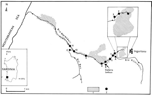
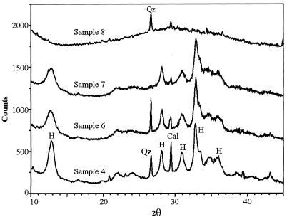
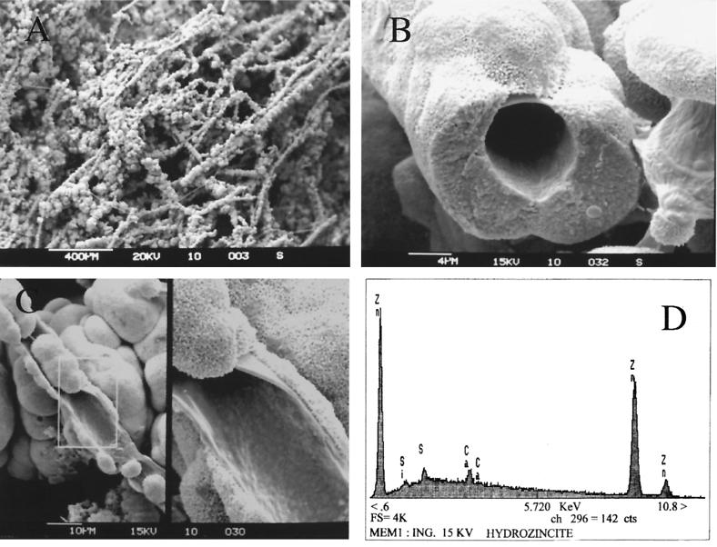
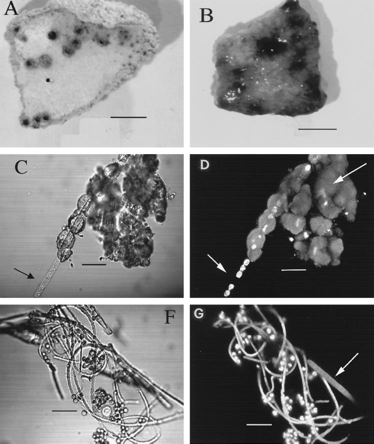
An iron-poor stream of nearly neutral pH polluted by mine tailings has been investigated for a natural phenomenon responsible for the polishing of heavy metals in mine wastewaters. A white mineralized mat, which was determined to be hydrozincite [Zn(5)(CO(3))(2)(OH)(6)] by X-ray diffraction analysis, was observed in the stream sediments mainly in spring. The precipitate shows a total organic matter residue of 10% dry weight and contains high concentrations of Pb, Cd, Ni, Cu, and other metals. Scanning electron microscopy analysis suggests that hydrozincite is mainly of biological origin. Dormant photosynthetic microorganisms have been retrieved from 1-year-old dry hydrozincite. The autofluorescent microorganisms were imaged by a scanning confocal laser microscope. A photosynthetic filamentous bacterium, classified as Scytonema sp. strain ING-1, was found associated with microalga Chlorella sp. strain SA1. This microbial community is responsible for the natural polishing of heavy metals in the water stream by coprecipitation with hydrozincite.
Figures
References
-
- Baldi F, Kukhar V P, Ulberg Z R. Bioconversion and removal of metals and radionuclides. In: Wild J R, editor. Perspectives in bioremediation. Amsterdam, The Netherlands: Kluver Academic Publishers; 1997. pp. 75–91.
-
- Baldi F, Minacci A, Saliot A, Mejanelle L, Mozetic O, Turk V, Malej A. Cell lysis and release of particulate polysaccharides in extensive marine mucillage assessed by lipid biomarkers and molecular probes. Mar Ecol Prog Ser. 1997;153:45–57.
-
- Buurman P, van Lagen B, Velthorst E J. Total or organic carbon and total nitrogen (elemental analyzer) In: Buurman P, van Lagen B, Velthorst E J, editors. Manual for soil and water analysis. Leiden, The Netherlands: Backhuys Publishers; 1996. pp. 11–16.
-
- Caboi R, Cidu R, Cristini A, Fanfani L, Massoli-Novelli R, Zuddas P. The abandoned Pb-Zn mine of Ingurtosu, Sardinia (Italy) Eng Geol. 1993;34:211–218.
-
- Caboi R, Cidu R, Fanfani L, Zuddas P. Abandoned mine sites: implications for water quality. In: Ciccu R, editor. Proceedings of the Fourth International Conference on Environmental Issues and Waste Management in Energy and Mineral Production. Vol. 2. Cagliari, Italy: Department of Geoengineering and Environmental Technologies, University of Cagliari; 1996. pp. 797–805.
Publication types
MeSH terms
Substances
LinkOut - more resources
Full Text Sources
Medical
