

Functional triads consisting of ryanodine receptors, Ca(2+) channels, and Ca(2+)-activated K(+) channels in bullfrog sympathetic neurons. Plastic modulation of action potential
- PMID: 11055998
- PMCID: PMC2229477
- DOI: 10.1085/jgp.116.5.697
Functional triads consisting of ryanodine receptors, Ca(2+) channels, and Ca(2+)-activated K(+) channels in bullfrog sympathetic neurons. Plastic modulation of action potential
Abstract
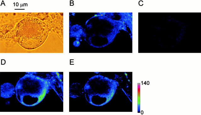
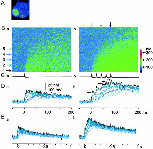
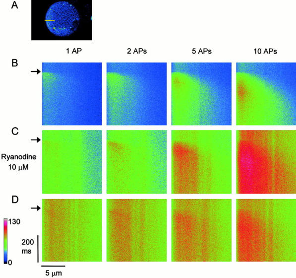
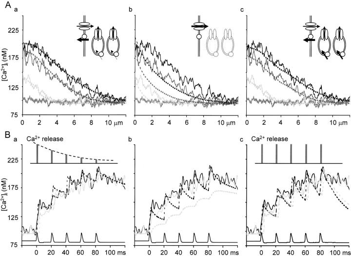
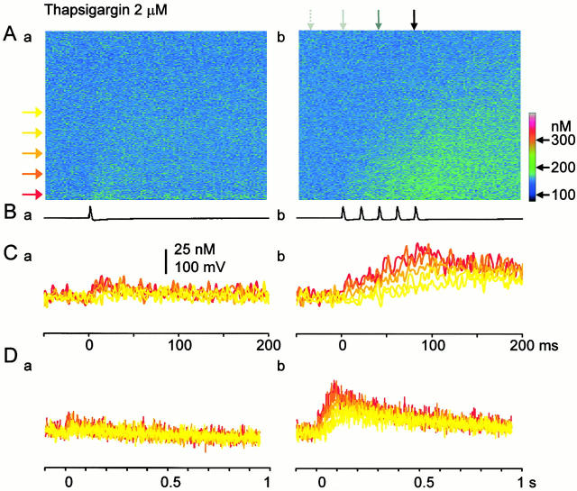
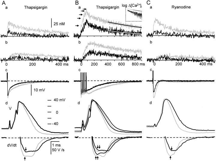
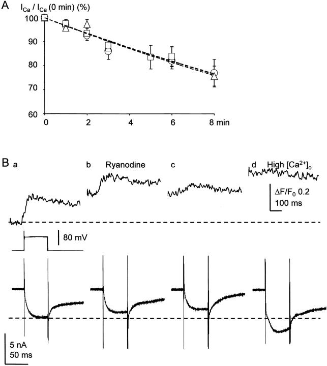
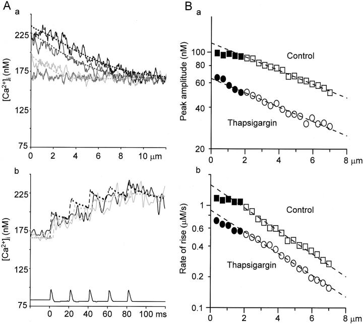
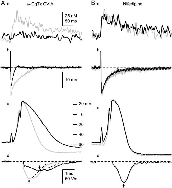
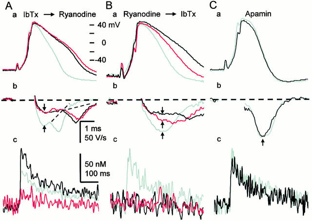
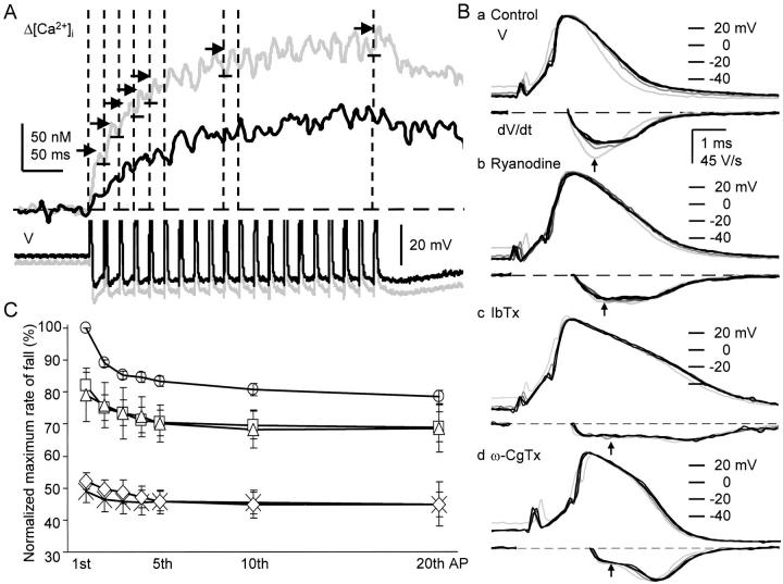
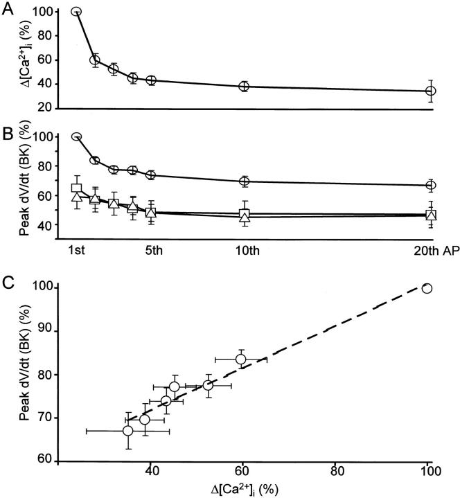
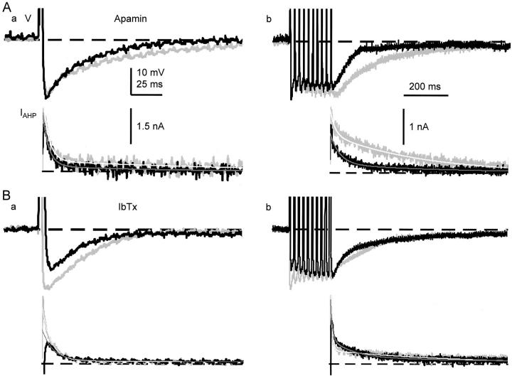
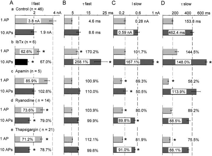
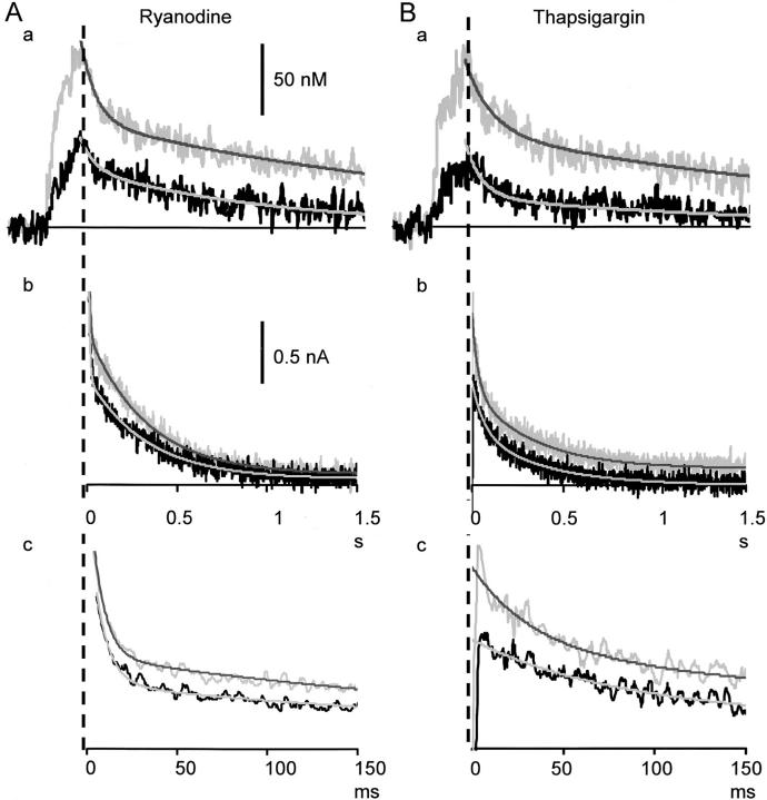
Fluorescent ryanodine revealed the distribution of ryanodine receptors in the submembrane cytoplasm (less than a few micrometers) of cultured bullfrog sympathetic ganglion cells. Rises in cytosolic Ca(2+) ([Ca(2+)](i)) elicited by single or repetitive action potentials (APs) propagated at a high speed (150 microm/s) in constant amplitude and rate of rise in the cytoplasm bearing ryanodine receptors, and then in the slower, waning manner in the deeper region. Ryanodine (10 microM), a ryanodine receptor blocker (and/or a half opener), or thapsigargin (1-2 microM), a Ca(2+)-pump blocker, or omega-conotoxin GVIA (omega-CgTx, 1 microM), a N-type Ca(2+) channel blocker, blocked the fast propagation, but did not affect the slower spread. Ca(2+) entry thus triggered the regenerative activation of Ca(2+)-induced Ca(2+) release (CICR) in the submembrane region, followed by buffered Ca(2+) diffusion in the deeper cytoplasm. Computer simulation assuming Ca(2+) release in the submembrane region reproduced the Ca(2+) dynamics. Ryanodine or thapsigargin decreased the rate of spike repolarization of an AP to 80%, but not in the presence of iberiotoxin (IbTx, 100 nM), a BK-type Ca(2+)-activated K(+) channel blocker, or omega-CgTx, both of which decreased the rate to 50%. The spike repolarization rate and the amplitude of a single AP-induced rise in [Ca(2+)](i) gradually decreased to a plateau during repetition of APs at 50 Hz, but reduced less in the presence of ryanodine or thapsigargin. The amplitude of each of the [Ca(2+)](i) rise correlated well with the reduction in the IbTx-sensitive component of spike repolarization. The apamin-sensitive SK-type Ca(2+)-activated K(+) current, underlying the afterhyperpolarization of APs, increased during repetitive APs, decayed faster than the accompanying rise in [Ca(2+)](i), and was suppressed by CICR blockers. Thus, ryanodine receptors form a functional triad with N-type Ca(2+) channels and BK channels, and a loose coupling with SK channels in bullfrog sympathetic neurons, plastically modulating AP.
Figures

Comment in
-
Functional interactions in Ca(2+) signaling over different time and distance scales.J Gen Physiol. 2000 Nov;116(5):691-6. doi: 10.1085/jgp.116.5.691. J Gen Physiol. 2000. PMID: 11055997 Free PMC article. No abstract available.
References
-
- Adams P.R., Constanti A., Brown D.A., Clark R.B. Intracellular Ca2+ activates a fast voltage-sensitive K+ current in vertebrate sympathetic neurones. Nature. 1982;296:746–749. - PubMed
-
- Akita T., Kuba K. Ca2+ release from submembrane and perinuclear Ca2+ stores are involved in action potential-induced Ca2+ transients and modulation of membrane excitability in cultured bullfrog sympathetic neurons Jpn. J. Physiol. 49Suppl.1999. S120a (Abstr.).
-
- Akita T., Kuba K. Ca2+ release from the submembrane and perinuclear Ca2+ stores induced by action potentials and its modulation of the cell membrane excitability in cultured sympathetic neurons Soc. Neurosci. Abstr 25 1999. 1999b (Abstr.).
-
- Allbritton N.L., Meyer T., Stryer L. Range of messenger action of calcium ion and inositol 1,4,5-trisphosphate. Science. 1992;258:1812–1815. - PubMed
Publication types
MeSH terms
Substances
LinkOut - more resources
Full Text Sources
Other Literature Sources
Miscellaneous
