. 2000;2(3):229-35.
doi: 10.1186/ar92. Epub 2000 Mar 1.

Apoptosis and p53 expression in rat adjuvant arthritis

Affiliations

Apoptosis and p53 expression in rat adjuvant arthritis

P P Tak et al. Arthritis Res. 2000.

Abstract

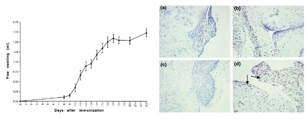
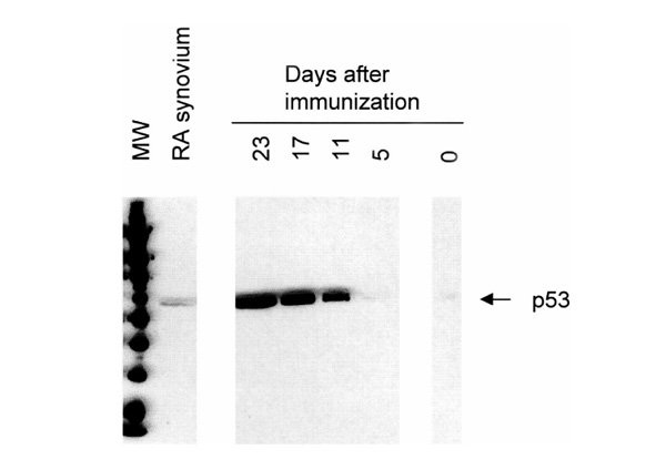
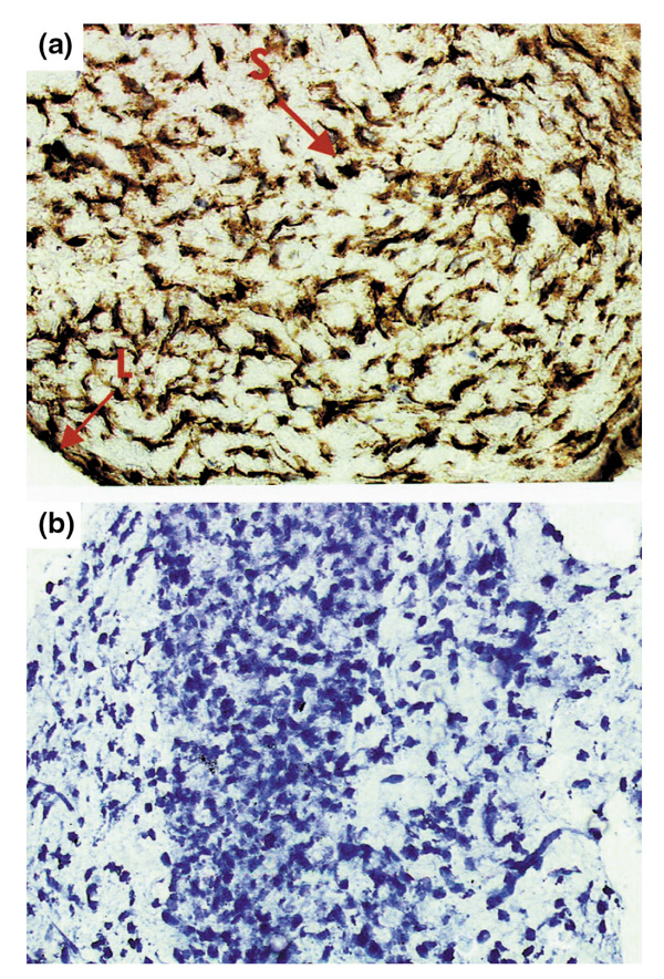
STATEMENT OF FINDINGS: The kinetics of apoptosis and the apoptosis-regulating gene p53 in adjuvant arthritis (AA) were investigated to assess the value of the AA rat model for testing apoptosis-inducing therapies. Very few terminal deoxynucleotidyl transferase-mediated deoxyuridine triphosphate (dUTP) nick end-labeling (TUNEL)-positive cells were detected during the early phases of AA, but on day 23 (chronic arthritis) the percentage of TUNEL-positive cells was significantly increased. Expression of p53 in synovial tissue gradually increased from days 5-23, which was markedly higher than p53 levels in rheumatoid arthritis (RA) synovium. Significant apoptosis only occurs late in rat AA and is concordant with marked p53 overexpression, making it useful model for testing proapoptotic therapies, but rat AA is not the best model for p53 gene therapy because dramatic p53 overexpression occurs in the latter stages of the disease.


Figures

Figure 1
Figure 1
(Left) Swelling of the left hind paws (mean ± standard error of the mean) in different phases of adjuvant arthritis (AA). Swelling usually starts around day 10 after immunization, followed by the development of accelerating arthritis (day 17) and chronic arthritis (day 23). (Right) Distribution of TUNEL-positive cells (red, indicated by arrows) in joints of AA rats on day 11 (a and c) and on day 23 (b and d). Very few TUNEL-positive cells were detected in normal rats or during the onset of arthritis, whereas the number of TUNEL-positive cells were significantly increased in the synovium (and also in cartilage) of rats with chronic AA. TUNEL method counterstrained with Mayer's hemalum. Original magnification 250x.
Figure 2
Figure 2
Western blot analysis showing immunoreactive p53 in pooled joint extracts of rats with AA and pooled synovial tissue samples of patients with RA. Expression of p53 gradually increased from days 0-23 in rat AA (see also Table 1). Overexpression on day 23 (173 arbitrary units) was markedly higher than p53 levels in RA synovium (32 arbitrary units).
Figure 3
Figure 3
Representative synovial tissue from a rat with adjuvant arthritis on day 23, showing marked p53 overexpression [(a), indicated by arrows]. Both cytoplasmic and nuclear staining was noted in the intimal lining layer (L) and in the synovial sublining (S). Staining was absent in the negative control section (b). Monostaining peroxidase technique with tyramine enhancement counterstained with Mayer's hemalum. Original magnification 400x.

Comment in

  • p53 in rheumatoid arthritis: friend or foe?
    Müller-Ladner U, Nishioka K. Müller-Ladner U, et al. Arthritis Res. 2000;2(3):175-8. doi: 10.1186/ar82. Epub 2000 Mar 31. Arthritis Res. 2000. PMID: 11094424 Free PMC article. Review.

References

    1. Firestein GS, Nguyen K, Aupperle KR, Yeo M, Zvaifler NJ. Apoptosis in rheumatoid arthritis: p53 overexpression in rheumatoid arthritis synovium. Am J Pathol. 1996;149:2143–2151. - PMC - PubMed
    1. Tak PP, Firestein GS. Apoptosis in rheumatoid arthritis. . Apoptosis and Inflammation Edited by Winkler JD Basel: Birkhauser Publishing Ltd. 1999:149–162.
    1. Firestein GS, Yeo M, Zvaifler NJ. Apoptosis in rheumatoid arthritis synovium. . J Clin Invest. 1995;96:1631–1638. - PMC - PubMed
    1. Nakajima T, Aono H, Hasunuma T, et al. Apoptosis and functional Fas antigen in rheumatoid arthritis synoviocytes. . Arthritis Rheum. 1995;38:485–491. - PubMed
    1. Tak PP, Zvaifler NJ, Green DR, Firestein GS. Rheumatoid arthritis and p53: how oxidative stress might alter the course of inflammatory diseases. . Immunol Today. 2000;21:78–82. - PubMed

Substances