

Heterogeneous nuclear ribonucleoproteins C1/C2 identified as autoantigens by biochemical and mass spectrometric methods
- PMID: 11056675
- PMCID: PMC17817
- DOI: 10.1186/ar119
Heterogeneous nuclear ribonucleoproteins C1/C2 identified as autoantigens by biochemical and mass spectrometric methods
Abstract
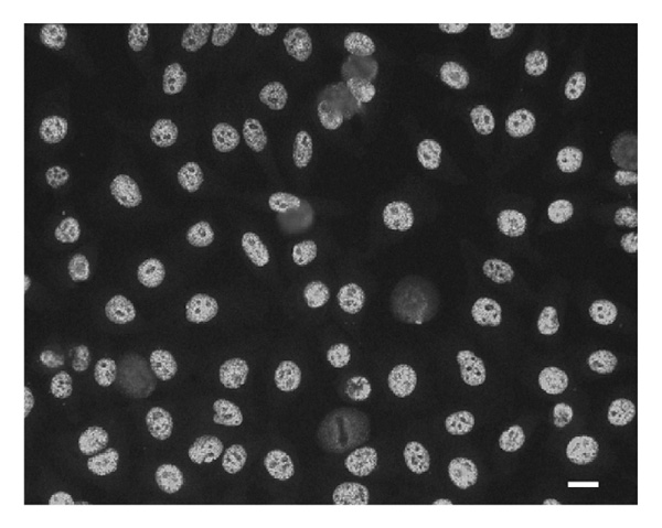
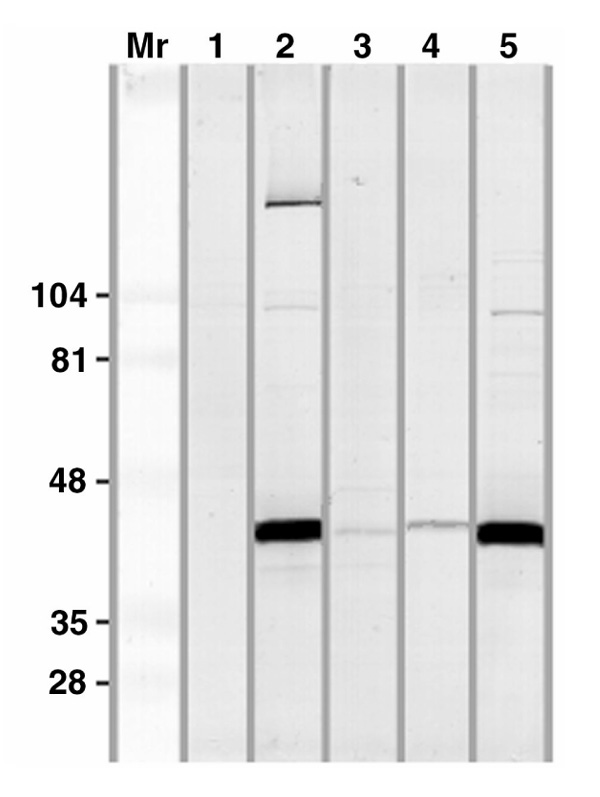
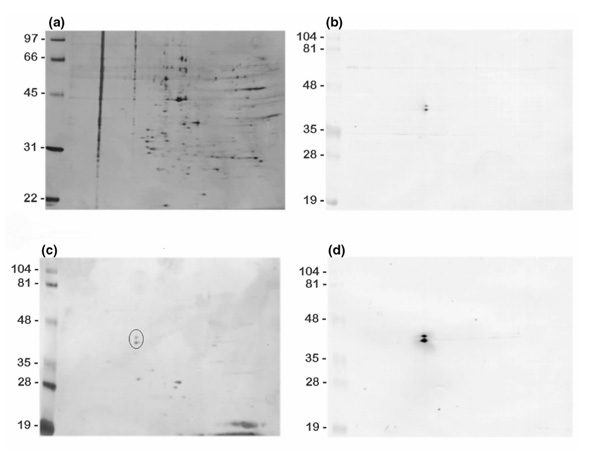
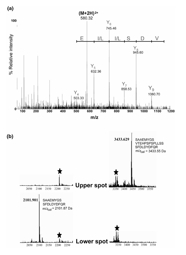
The antigenic specificity of an unusual antinuclear antibody pattern in three patient sera was identified after separating HeLa-cell nuclear extracts by two-dimensional (2D) gel electrophoresis and localizing the antigens by immunoblotting with patient serum. Protein spots were excised from the 2D gel and their contents were analyzed by matrix-assisted laser desorption-ionization (MALDI) or nanoelectrospray ionization time-of-flight (TOF) tandem mass spectrometry (MS) after in-gel digestion with trypsin. A database search identified the proteins as the C1 and C2 heterogeneous nuclear ribonucleoproteins. The clinical spectrum of patients with these autoantibodies includes arthritis, psoriasis, myositis, and scleroderma. None of 59 patients with rheumatoid arthritis, 19 with polymyositis, 33 with scleroderma, and 10 with psoriatic arthritis had similar antibodies. High-resolution protein-separation methods and mass-spectrometric peptide mapping in combination with database searches are powerful tools in the identification of novel autoantigen specificities.
Figures
Similar articles
-
Decreased brain levels of Lupus La protein and increased U5 small ribonucleoprotein-specific 40 kDa protein in fetal Down syndrome.Cell Mol Biol (Noisy-le-grand). 2003 Jul;49(5):733-8. Cell Mol Biol (Noisy-le-grand). 2003. PMID: 14528909
-
Two-dimensional electrophoresis and mass spectrometry identification of proteins bound by a murine monoclonal anti-cardiolipin antibody: a powerful technique to characterize the cross-reactivity of a single autoantibody.Electrophoresis. 2000 Jul;21(12):2531-9. doi: 10.1002/1522-2683(20000701)21:12<2531::AID-ELPS2531>3.0.CO;2-E. Electrophoresis. 2000. PMID: 10939468
-
Multiple specificities of autoantibodies against hnRNP A/B proteins in systemic rheumatic diseases and hnRNP L as an associated novel autoantigen.Autoimmunity. 2007 May;40(3):223-33. doi: 10.1080/08916930701352357. Autoimmunity. 2007. PMID: 17453722
-
Autoantibodies to heterogeneous nuclear ribonucleoproteins.Autoimmunity. 2005 Feb;38(1):25-32. doi: 10.1080/08916930400022590. Autoimmunity. 2005. PMID: 15804702 Review.
-
Update on autoantibodies to intracellular antigens in systemic rheumatic diseases.Clin Lab Med. 1992 Mar;12(1):1-23. Clin Lab Med. 1992. PMID: 1563236 Review.
Cited by
-
ADAR and hnRNPC deficiency synergize in activating endogenous dsRNA-induced type I IFN responses.J Exp Med. 2021 Sep 6;218(9):e20201833. doi: 10.1084/jem.20201833. Epub 2021 Jul 23. J Exp Med. 2021. PMID: 34297039 Free PMC article.
-
An Autoantigen Profile of Human A549 Lung Cells Reveals Viral and Host Etiologic Molecular Attributes of Autoimmunity in COVID-19.bioRxiv [Preprint]. 2021 Feb 22:2021.02.21.432171. doi: 10.1101/2021.02.21.432171. bioRxiv. 2021. Update in: J Autoimmun. 2021 Jun;120:102644. doi: 10.1016/j.jaut.2021.102644. PMID: 33655248 Free PMC article. Updated. Preprint.
-
A Master Autoantigen-ome Links Alternative Splicing, Female Predilection, and COVID-19 to Autoimmune Diseases.bioRxiv [Preprint]. 2021 Aug 4:2021.07.30.454526. doi: 10.1101/2021.07.30.454526. bioRxiv. 2021. Update in: J Transl Autoimmun. 2022;5:100147. doi: 10.1016/j.jtauto.2022.100147. PMID: 34373855 Free PMC article. Updated. Preprint.
-
Protein disulfide isomerases are antibody targets during immune-mediated tumor destruction.Blood. 2009 Feb 19;113(8):1681-8. doi: 10.1182/blood-2007-09-114157. Epub 2008 Nov 13. Blood. 2009. PMID: 19008459 Free PMC article. Clinical Trial.
-
An Autoantigen Atlas From Human Lung HFL1 Cells Offers Clues to Neurological and Diverse Autoimmune Manifestations of COVID-19.Front Immunol. 2022 Mar 24;13:831849. doi: 10.3389/fimmu.2022.831849. eCollection 2022. Front Immunol. 2022. PMID: 35401574 Free PMC article.
References
-
- Hassfeld W, Steiner G, Studnicka-Benke A, Skriner K, Graninger W, Fischer I, Smolen JS. Autoimmune response to the spliceosome: an immunologic link between rheumatoid arthritis, mixed connective tissue disease, and systemic lupus erythematosus. Arthritis Rheum. 1995;6:777–785. - PubMed
-
- Zimmermann C, Steiner G, Skriner K, Hassfeld W, Petera P, Smolen JS. The concurrence of rheumatoid arthritis and limited systemic sclerosis: clinical and serologic characteristics of an overlap syndrome. . Arthritis Rheum. 1998;41:1938–1945. - PubMed
-
- Steiner G, Skriner K, Smolen JS. Autoantibodies to the A/B proteins of the heterogeneous nuclear ribonucleoprotein complex: novel tools for the diagnosis of rheumatic diseases. Int Arch Allergy Appl Immunol. 1996;111:314–319. - PubMed
-
- Stanek D, Vencovský J, Kafková J, Raska I. Heterogenous nuclear RNP C1 and C2 core proteins are targets for an autoantibody found in the serum of a patient with systemic sclerosis and psoriatic arthritis. . Arthritis Rheum. 1997;40:2172–2177. - PubMed
-
- Nawrocki A, Larsen MR, Podtelejnikov AV, Jensen ON, Mann M, Roepstorff P, Görg A, Fey SJ, Mose-Larsen P. Correlation of acidic and basic carrier ampholyte and immobilized pH gradient two-dimensional gel electrophoresis patterns based on mass spectrometric protein identification. . Electrophoresis. 1998;19:1024–1035. - PubMed
Publication types
MeSH terms
Substances
LinkOut - more resources
Full Text Sources
Miscellaneous
