. 2000;2(6):477-88.
doi: 10.1186/ar130. Epub 2000 Aug 31.

Mesenchymal precursor cells in the blood of normal individuals

Affiliations

Mesenchymal precursor cells in the blood of normal individuals

N J Zvaifler et al. Arthritis Res. 2000.

Abstract

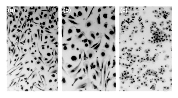
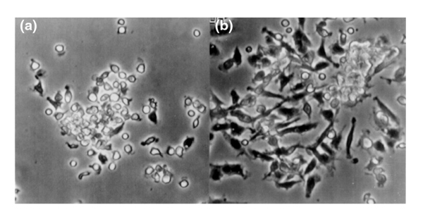
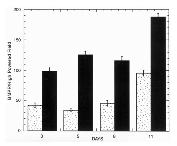
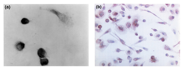
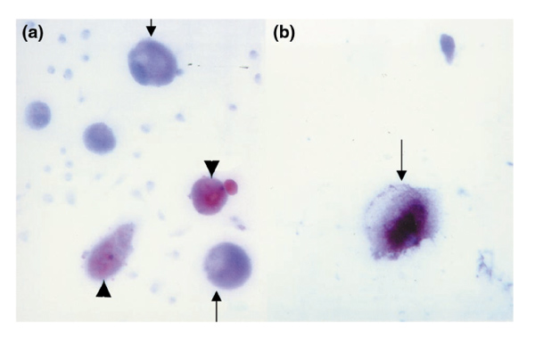
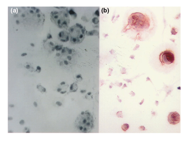
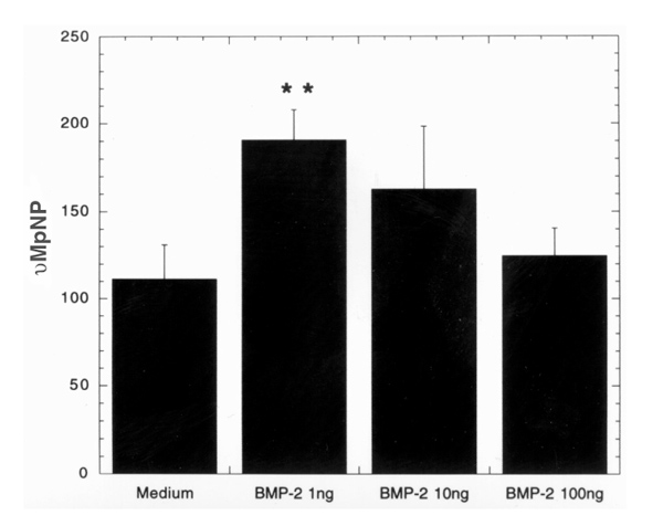
STATEMENT OF FINDINGS: Mesenchymal precursor cells found in the blood (BMPCs) of normal persons adhere to plastic and glass and proliferate logarithmically in DMEM-20% fetal calf serum (FCS) without growth factors. They form cells with fibroblast-like and stromal morphology, which is not affected by eliminating CD34, CD3, or CD14 cells. Osteogenic supplements (dexamethasone, ascorbic acid, and beta-glycerophosphate) added to the culture inhibited fibroblast formation, and BMPCs assumed the cuboidal shape of osteoblasts. After 5 days in supplemented medium, the elutriated cells displayed alkaline phosphatase (AP), and the addition of bone morphogenetic protein (BMP)2 (1 ng) doubled AP production (P < 0.04). Two weeks later, 30% of the cells were very large and reacted with anti-osteocalcin antibody. The same cultures also contained sudanophlic adipocytes and multinucleated giant cells that stained for tartrate-resistant acid phosphatase (TRAP) and vitronectin receptors. Cultured BMPCs immunostain with antibodies to vimentin, type I collagen, and BMP receptors, heterodimeric structures expressed on mesenchymal lineage cells. In addition, BMPCs stain with anti-CD105 (endoglin), a putative marker for bone-marrow mesenchymal stem cells (MSCs).


Figures

Figure 1
Figure 1
Appearance of cells isolated from a BMPC-rich elutriation fraction of healthy human blood and cultured in DMEM-20% FCS for 8 days (a, b). At this time, the predominant cells consist of both fibroblast-like cells with a central nucleus, filmy cytoplasm, and adherent pseudopods, and large, round cells with a thin, adherent cytoplasm and a round, central nucleus. Hematoxylin staining. (a) ×200; (b) ×400. (c) The appearance of cells isolated from the same BMPC-rich elutriation fraction of healthy human blood and cultured in DMEM-20% FCS supplemented at the initiation of culture with dexamethasone (10-7mol), ascorbic-acid-2-phosphate (0.05 mmol), and β-glycerophosphate (10 mmol) for 8 days. At this time, all the cells have a round or cuboidal morphology with a centrally placed nucleus. ×200. Compare with the nonsupplemented cultures in (a).
Figure 2
Figure 2
Time-lapse video cinematography of a BMPC-rich elutriation fraction cultured in DMEM-20% FCS observed by phase-contrast microscopy. At day 2 (a), there are small, round cells in clusters and a few cells with pseudopods. By day 6 (b), there are many large, fibroblast-like cells and large, round, stromal cells arising from a cluster of small, round cells.
Figure 3
Figure 3
BMPC-rich elutriation fraction from healthy human blood cultured in DMEM-20% FCS. Cells were fixed, stained with anti-BMPR 1A antibody (see Methods), and analyzed on days 3, 5, 8, and 11. BMPCs were determined by morphology (fibroblast-like [hatched bars] or big cells [filled bars]) and immunoperoxidase staining. Results are presented as the means of total number of cells in six individual images. ×400. Computer image analysis (AnalySIS).
Figure 4
Figure 4
BMPC-rich elutriation fraction cultured on a sterile glass slide in DMEM-20% FCS and allowed to proliferate. (a) On day 7, the slide was washed and the cells were fixed and stained with anti-BMPR Type 1A antibody. In the background are many unstained, small, mononuclear cells. Two types of cell are stained: one looks like a fibroblast, and the other is a large, round cell with an adherent cytoplasm. (b) A similar culture on day 12, stained with anti-endoglin (CD105) antibody. The cytoplasm of almost all the large mesenchymal cells shows stippled staining with perinuclear accentuation.
Figure 5
Figure 5
BMPC-rich elutriation fraction cultured in DMEM-20% FCS supplemented with dexamethasone, ascorbic acid, and β-glycerophosphate (as described in Fig. 1). After 20 days, about one-third of the cells are very large. Their cytoplasm stains with an anti-calcitonin antibody. Immunoperoxidase staining is also observed in the matrix formed around some of the largest cells. In the same supplemented cultures are large cells containing neutral lipid (Sudan IV; red stain). (a) Shows both anti-calcitonin-stained cells (arrows) and sudanophilic cells (arrowheads) in the same supplemented cultures (×250) and (b) is at higher magnification (×400) showing a cell and its surrounding matrix stained with an anticalcitonin antibody (arrow).
Figure 6
Figure 6
BMPC-rich elutriation fraction cultured in DMEM-20% FCS supplemented with dexamethasone, ascorbic acid, and β-glycerophosphate (as described in Fig. 1) and examined daily thereafter. (a) By day 7 there are many large, multinucleated cells (phase-contrast microscopy). (b) These cells stain with an anti-vitronectin receptor antibody (×250).
Figure 7
Figure 7
BMPC-rich elutriation fractions (n = 4) cultured in DMEM-20% FCS with varying concentrations of BMP2 for 5 days. Supernatants were collected and analyzed for alkaline phosphatase activity (AP; the release of pNP is a measure of AP activity). The lowest concentration of BMP2 (1 ng/ml) caused a significant increase in AP activity. **P > 0.004.
Figure 8
Figure 8
RT-PCR analysis for expression of mRNA for (a) the chemokine SDF-1 and (b) the housekeeping gene GA3PD in cultured BMPCs (GA42, GA43), and a rheumatoid arthritis synovial fibroblast line (RA505 passage 4). Note the similar RT-PCR fragment size in the samples to that in an SDF-1 plasmid (lane 5) included as a positive control. Conditions for RT-PCR and the primers used are described in the methods section.

References

    1. Caplan AI. Mesenchymal stem cells. J Orthop Res. 1991;9:641–650. - PubMed
    1. Friedenstein AJ, Gorskaja JF, Kulagina NN. Precursor cells of mechanocytes. ExpHematol. 1976;4:267–274. - PubMed
    1. Bruder SP, Jaiswal N, Haynesworth SE. Growth kinetics, self-renewal, and the osteogenic potential of purified human mesenchymal stem cells during extensive subcultivation and following cryopreservation. . J Cell Biochem. 1997;64:278–294. - PubMed
    1. Barry FP, Boynton RE, Haynesworth S, Murphy JM, Zaia J. The monoclonal antibody SH-2, raised against human mesenchymal stem cells, recognizes an epitope on endoglin (CD105). Biochem Biophys Res Commun. 1999;265:134–139. - PubMed
    1. Rosen V. Signaling pathways in skeletal formation. A role for BMP receptors. In Ann NY Acad Sci No 785: Molecular and Developmental Biology of Cartilage Edited by de Crombrugghe B. 1996. pp. 56–59. - PubMed

Publication types