. 2000;2(3):222-35.
doi: 10.1186/bcr57. Epub 2000 Mar 7.

A novel cell culture model for studying differentiation and apoptosis in the mouse mammary gland

Affiliations

A novel cell culture model for studying differentiation and apoptosis in the mouse mammary gland

K E Gordon et al. Breast Cancer Res. 2000.

Abstract

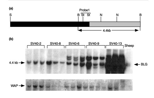
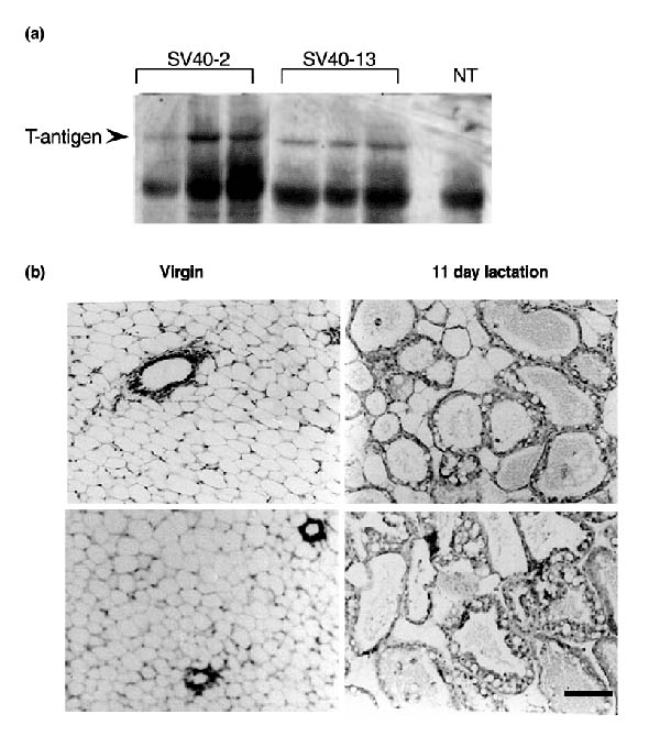
Background: This paper describes the derivation and characterization of a novel, conditionally immortal mammary epithelial cell line named KIM-2. These cells were derived from mid-pregnant mammary glands of a mouse harbouring one to two copies of a transgene comprised of the ovine beta-lactoglobulin milk protein gene promoter, driving expression of a temperature-sensitive variant of simian virus-40 (SV40) large T antigen (T-Ag).

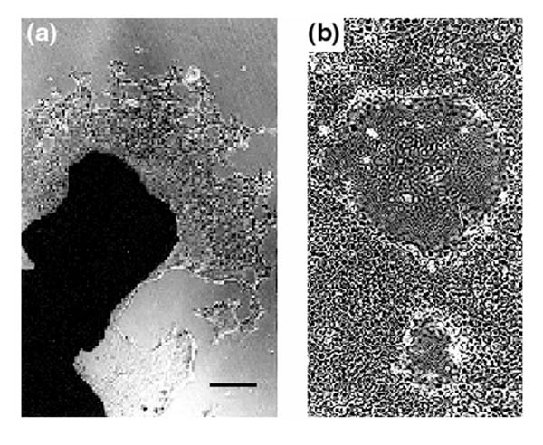
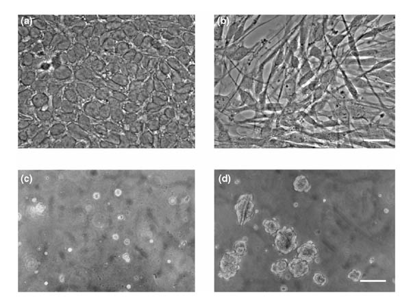
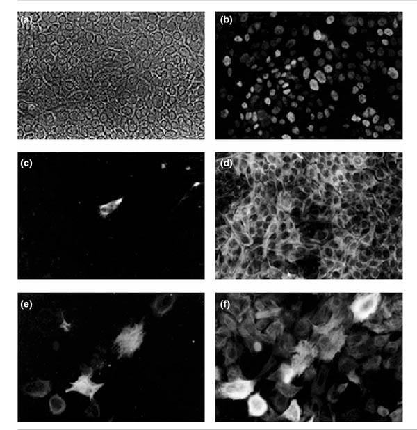
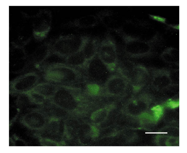
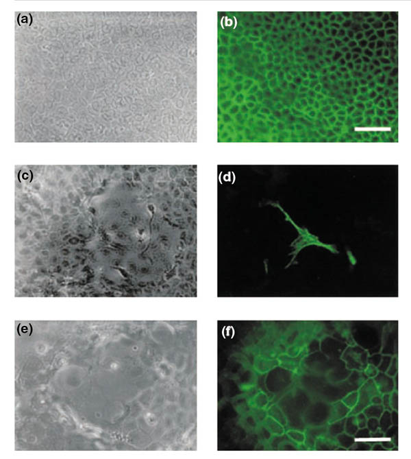
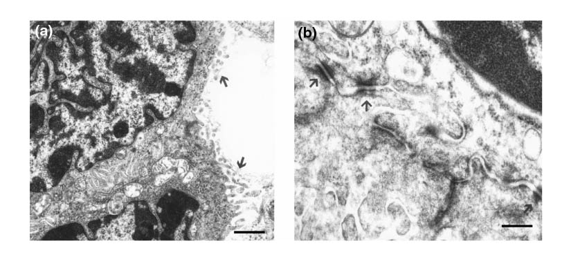
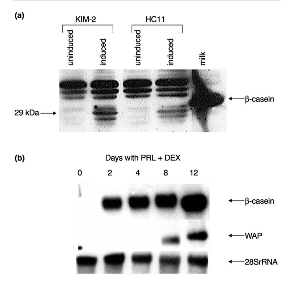
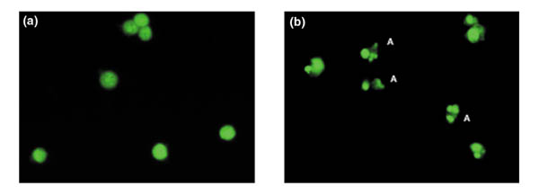
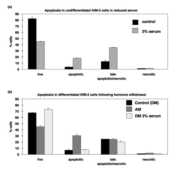
Results: KIM-2 cells have a characteristic luminal epithelial cell morphology and a stable, nontransformed phenotype at the semipermissive temperature of 37 degrees C. In contrast, at the permissive temperature of 33 degrees C the cells have an elongated spindle-like morphology and become transformed after prolonged culture. Differentiation of KIM-2 cells at 37 degrees C, in response to lactogenic hormones, results in the formation of polarized dome-like structures with tight junctions. This is accompanied by expression of the milk protein genes that encode beta-casein and whey acidic protein (WAP), and activation of the prolactin signalling molecule, signal transducer and activator of transcription (STAT)5. Fully differentiated KIM-2 cultures at 37 degrees C become dependent on lactogenic hormones for survival and undergo extensive apoptosis upon hormone withdrawal, as indicated by nuclear morphology and flow cytometric analysis. KIM-2 cells can be genetically modified by stable transfection and clonal lines isolated that retain the characteristics of untransfected cells.

Conclusion: KIM-2 cells are a valuable addition, therefore, to currently available lines of mammary epithelial cells. Their capacity for extensive differentiation in the absence of exogenously added basement membrane, and ability to undergo apoptosis in response to physiological signals will provide an invaluable model system for the study of signal transduction pathways and transcriptional regulatory mechanisms that control differentiation and involution in the mammary gland.


Figures

Figure 1
Figure 1
BLG-tsA58 transgenic mice. (a) Scheme of vector construct; 4.2 kb of the ovine BLG promoter including the noncoding exon 1 was fused to the 'enhancerless' SV40 T-Ag temperature sensitive mutant tsA58. The restriction sites shown are Sall (S), BamHI (B), Stul (St), and Ndel (N). The EcoRV and Bgll sites were destroyed during the construction of this vector. (b) The transgene copy number in the five surviving transgenic lines was estimated by Southern blotting. Digested genomic liver DNA (10 μg) from three sisters in each of the lines was analyzed with sheep genomic liver DNA used as a single copy control. The blot was hybridized with probe 1, and stripped and rehybridized with a WAP probe as a loading control.
Figure 2
Figure 2
Expression of T-Ag in the mammary glands of transgenic mice harbouring one to two copies (line SV40-2) or about 10 copies (line SV40-13) of the BLG-tsA58 transgene. (a) Protein extracts (20 μg) were prepared from day 11 lactation mammary tissue from three age-matched sisters from each line and a nontransgenic control mouse. The extracts were separated by sodium dodecyl sulphate-polyacrylamide gel electrophoresis, electroblotted onto nitrocellulose and probed with an anti-T-Ag antibody, DP-02. The nonspecific band is also seen in the control sample. (b) Morphological comparison of haematoxylin and eosin stained sections of virgin and 11-day lactation mammary glands from transgenic line SV40-2 (top) and a nontransgenic control (bottom). Histologically, the transgenic glands were indistinguishable from control glands. Scale bar 100 μm.
Figure 3
Figure 3
Derivation of primary cultures from mammary outgrowths. (a) Explants isolated from pregnant mammary tissue from transgenic line SV40-2 developed outgrowths after 2 weeks in culture at 37°C on type 1 collagen. The typical cobblestone morphology that is characteristic of epithelial cells was observed in these outgrowths by phase contrast microscopy. (b) After 14 days in culture the explants were removed and the primary cultures maintained for a further 7-14 days. Dome structures formed during this time period. Scale bar 250 μm.
Figure 4
Figure 4
Effects of growth temperature on morphology and anchorage independent growth of KIM-2. (a) Cells isolated and grown at 37°C showing the typical cuboidal epithelial morphology. (b) Cells isolated and grown at 33°C have a spindle-like mesenchymal morphology. Scale bar for (a) and (b) 50 μm. (c) Cells, isolated and grown at 37°C, seeded in soft agar and cultured for 12 days. Cells do not form colonies. (d) Cells, isolated and grown at 33°C, seeded in soft agar and cultured for 12 days at 37°C showing the formation of colonies. Scale bar for (c) and (d) 10 μm.
Figure 5
Figure 5
Immunocytochemical analysis of KIM-2 cells cultures at 37°C. (a) Phase contrast. (b) The same field showing nuclear staining with a T-Ag-specific antibody (Pab 419). (c) Staining with a vimentin monoclonal antibody (Vim-13.2) was detected in some fields. (d) Strong positive staining with a keratin 18-specific monoclonal antibody (LE61), a luminal epithelial marker. Scale bar for (a)-(d) 50 μm. (e) Staining with a smooth muscle actin monoclonal antibody, a myoepithelial marker. (f) Positive staining with a keratin 14-specific antibody (LL002). The epitope recognized by this antibody is often deregulated in culture. Scale bar for (e) and (f) 25 μm.
Figure 6
Figure 6
Deposition of laminin by KIM-2 cells. A confluent monolayer of undifferentiated KIM-2 cells was stained with an antibody to laminin and detected by FITC-conjugated secondary antibody. Control slides with secondary antibody alone were completely blank. Scale bar 25 μm.
Figure 7
Figure 7
Analysis of junction formation and differentiation of KIM-2 cells. (a) Phase contrast. (b) Detection of E-cadherin on surface of confluent monolayer of cells. Scale bar for (a) and (b) 50 μm. (c) Phase contrast of differentiated cells and (d) detection of myoepithelial cell on surface of dome with an antibody to smooth muscle α-actin. Scale bar for (c) and (d) 50 μm. (e) Phase contrast of dome and (f) detection of tight junctions with antibody to zonula occluden-1. Scale bar for (e) and (f) 25 μm.
Figure 8
Figure 8
Electron microscopy of differentiated KIM-2 cells. (a) Microvilli (arrowed) on the apical surface of cells and showing the interdigitated cytoplasm at the boundary between two cells. Scale bar 2 μm. (b) Higher magnification showing the presence of desmosomes (arrowed). Scale bar 0.5 μm.
Figure 9
Figure 9
Analysis of the differentiation capacity of KIM-2 and HC11 cells in response to lactogenic hormones on tissue culture plastic. (a) Endogenous β-casein expression was analyzed in KIM-2 and HC11 cells by western blot. Total cell lysates (20 μg) were prepared from KIM-2 cells or HC11 cells grown to confluence and induced with the lactogenic hormone cocktail of insulin, prolactin and dexamethasone for 4 days at 37°C. Uninduced cells were grown to confluence in growth medium. A defatted mouse milk sample was used as a positive control for the murine β-casein polyclonal antibody (diluted 1:10 000). Intracellular β-casein appeared to be a doublet at approximately 29 kDa in extracts from both cell lines, whereas a 32kDa secreted β-casein was detected in defatted milk. This size discrepancy can be accounted for by differences in phosphorylation states between intracellular and secreted forms of the protein. (b) WAP and β-casein expression were analyzed in KIM-2 cells by northern blot. Total RNA (20 μg) was prepared from KIM-2 cells grown to confluence at 37°C and harvested or induced with lactogenic hormones for the time period indicated. WAP mRNA was detected after 8 days exposure to lactogenic hormones. Figures are representative of three separate experiments, although induction of WAP expression was variable and occurred between 4 and 8 days after hormone treatment in different experiments.
Figure 10
Figure 10
Analysis of STAT5 activation by electrophoretic mobility shift assay in KIM-2 cells stimulated with prolactin. Nuclear extracts were prepared from confluent KIM-2 cultures stimulated with prolactin, in the absence of EGF, for the time periods indicated. The track labelled Mamm is an extract from a day 2 lactation mouse mammary gland. Uninduced control cells were maintained in growth medium. Extracts (4 μg protein) were incubated with a radioactively labelled DNA probe containing the highest affinity ovine BLG STAT5 binding site [34] and complexes resolved on a nondenaturing 6% acrylamide gel. A representative of more than three experiments is shown.
Figure 11
Figure 11
Induction of apoptosis in KIM-2 cells. KIM-2 cells were cultured in reduced serum and the absence of additional growth factors for 24 h, inducing apoptosis in approximately 40% of the cells. (a) Control and (b) dying KIM-2 cells were stained with acridine orange and visualized by fluorescence microscopy. Examples of cells exhibiting classical apoptotic morphology are indicated by the letter 'A'.
Figure 12
Figure 12
Analysis of apoptosis by flow cytometry. (a) Cells were induced to undergo apoptosis by culturing in reduced serum (3%) and the absence of additional growth factors for 24 h. (b) Cells that had been cultured in differentiation medium (DM) for 12 days were induced to undergo apoptosis by removal of prolactin, insulin and dexamethasone (AM) for 17 h. Control and dying cells were stained with annexin V and propidium iodide (PI) and analyzed by flow cytometry. PI enters only cells with damaged membranes (late apoptosis or necrosis). Four populations of cells were identified: live (no stain), apoptotic (annexin V only), late apoptotic/necrotic (annexin V +PI) and necrotic (PI) only. The percentage of cells in each population is shown as the mean of three experiments with the standard error of the mean.

Similar articles

Cited by

References

    1. Topper YJ, Freeman CS. Multiple hormone interactions in the developmental biology of the mammary gland. Physiol Rev. 1980;60:1049–1056. - PubMed
    1. Levine JF, Stockdale FE. Cell-cell interactions promote mammary epithelial cell differentiation. J Cell Biol. 1985;100:1415–1422. - PMC - PubMed
    1. Li M, Aggeler J, Farson DA, et al. Influence of reconstituted basement membrane and its components on casein expression and secretion in mouse mammary epithelial cells. Proc Natl Acad Sci USA. 1987;84:136–140. - PMC - PubMed
    1. Reichman E, Ball R, Groner B, Friis RR. New mammary epithelial and fibroblastic cell clones in coculture form structures competent to differentiate functionally. J Cell Biol. 1989;108:1127–1138. - PMC - PubMed
    1. Danielson KG, Oborn CJ, Durban EM, Butel JS, Medina D. Epithelial mouse mammary cell line exhibiting normal morphogenesis in vivo and functional differentiation in vitro. Proc Natl Acad Sci USA. 1984;81:3756–3760. - PMC - PubMed

Publication types

MeSH terms

LinkOut - more resources