Review
. 2000 Nov 1;28(21):4051-8.
doi: 10.1093/nar/28.21.4051.

Structural and mechanistic conservation in DNA ligases

Affiliations
Review

Structural and mechanistic conservation in DNA ligases

A J Doherty et al. Nucleic Acids Res. .

Abstract

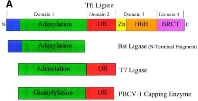
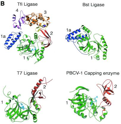
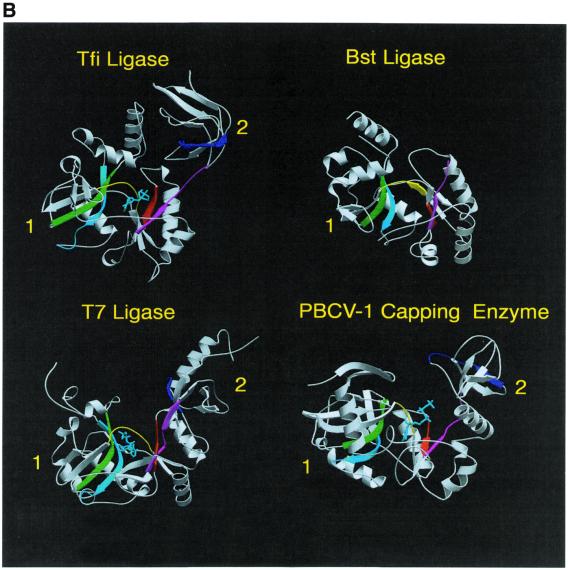
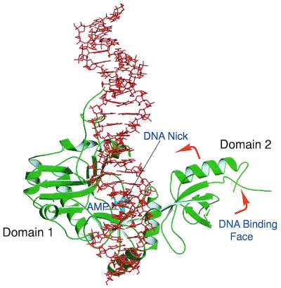
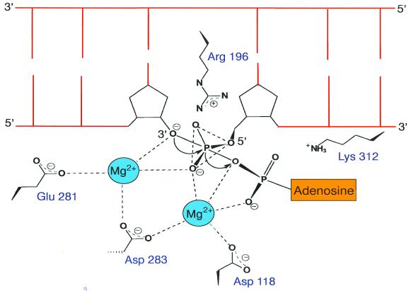
DNA ligases are enzymes required for the repair, replication and recombination of DNA. DNA ligases catalyse the formation of phosphodiester bonds at single-strand breaks in double-stranded DNA. Despite their occurrence in all organisms, DNA ligases show a wide diversity of amino acid sequences, molecular sizes and properties. The enzymes fall into two groups based on their cofactor specificity, those requiring NAD(+) for activity and those requiring ATP. The eukaryotic, viral and archael bacteria encoded enzymes all require ATP. NAD(+)-requiring DNA ligases have only been found in prokaryotic organisms. Recently, the crystal structures of a number of DNA ligases have been reported. It is the purpose of this review to summarise the current knowledge of the structure and catalytic mechanism of DNA ligases.


Figures

Figure 1
Figure 1
Mechanisms of DNA ligation. This figure shows the partial reactions catalysed by ATP- and NAD+-dependent DNA ligases. The structure of the phosphoramidate intermediate common to both reactions is also illustrated.
Figure 2
Figure 2
Domain structure of DNA ligases. (A) Schematic representation of the domain architecture of the known DNA ligase structures. The domains are colour coded: subdomain 1a, blue; subdomain 1b (adenylation), green; domain 2 (oligo-binding), red; subdomain 3a (zinc finger), yellow; subdomain 3b (helix–hairpin–helix), orange; domain 4 (BRCA1 C-terminus), pink. (B) A ribbon diagram representation of the structures of the NAD+-dependent ligases encoded by T.filiformis (Tfi) and B.stearothermophilus (Bst) (N-terminal domain only), the ATP-dependent DNA ligase of bacteriophage T7 (T7) and the capping enzyme encoded by Chlorella virus PBCV-1. The conserved structural domains have been numbered and coloured as in (A) to allow for ease of identification in the different structures.
Figure 2
Figure 2
Domain structure of DNA ligases. (A) Schematic representation of the domain architecture of the known DNA ligase structures. The domains are colour coded: subdomain 1a, blue; subdomain 1b (adenylation), green; domain 2 (oligo-binding), red; subdomain 3a (zinc finger), yellow; subdomain 3b (helix–hairpin–helix), orange; domain 4 (BRCA1 C-terminus), pink. (B) A ribbon diagram representation of the structures of the NAD+-dependent ligases encoded by T.filiformis (Tfi) and B.stearothermophilus (Bst) (N-terminal domain only), the ATP-dependent DNA ligase of bacteriophage T7 (T7) and the capping enzyme encoded by Chlorella virus PBCV-1. The conserved structural domains have been numbered and coloured as in (A) to allow for ease of identification in the different structures.
Figure 3
Figure 3
(A) Sequence motifs conserved in DNA ligases and RNA capping enzymes. Conserved sequence elements define a superfamily of covalent nucleotidyl transferases. Six sequence elements, designated motifs I, III, IIIa, IV, V and VI, are conserved in NAD+- and ATP-dependent DNA ligases and guanylyl transferases as shown. The alignment includes the NAD+-dependent ligases (Lig) encoded by T.filiformis (Tfi), B.stearothermophilus (Bst), E.coli (Eco) and T.thermophilus (Tth). Below these are aligned sequences for the ATP-dependent DNA ligases (Lig) of bacteriophage T7 (BT7), vaccinia virus (Vac), Saccharomyces cerevisiae (Sce), Schizosaccharomyces pombe (Spo) and human ligases I (Hu1), 3 (Hu3) and 4 (Hu4). The alignment also contains the amino acid sequences for capping enzymes (CE) encoded by Chlorella virus PBCV-1 (ChV), S.cerevisiae, S.pombe, Candida albicans (Cal), African swine fever virus (ASF), vaccinia virus and Caenorhabditis elegans (Cel). The numbers of amino acid residues separating the motifs are indicated. The active site lysine is shown in red. (B) The location of the conserved sequence motifs as described in (A) are indicated by the corresponding colours in the ribbon diagrams of the crystal structures of the Tfi, Bst and T7 DNA ligases and Chlorella virus RNA capping enzyme. The bound nucleotides are shown in cyan.
Figure 3
Figure 3
(A) Sequence motifs conserved in DNA ligases and RNA capping enzymes. Conserved sequence elements define a superfamily of covalent nucleotidyl transferases. Six sequence elements, designated motifs I, III, IIIa, IV, V and VI, are conserved in NAD+- and ATP-dependent DNA ligases and guanylyl transferases as shown. The alignment includes the NAD+-dependent ligases (Lig) encoded by T.filiformis (Tfi), B.stearothermophilus (Bst), E.coli (Eco) and T.thermophilus (Tth). Below these are aligned sequences for the ATP-dependent DNA ligases (Lig) of bacteriophage T7 (BT7), vaccinia virus (Vac), Saccharomyces cerevisiae (Sce), Schizosaccharomyces pombe (Spo) and human ligases I (Hu1), 3 (Hu3) and 4 (Hu4). The alignment also contains the amino acid sequences for capping enzymes (CE) encoded by Chlorella virus PBCV-1 (ChV), S.cerevisiae, S.pombe, Candida albicans (Cal), African swine fever virus (ASF), vaccinia virus and Caenorhabditis elegans (Cel). The numbers of amino acid residues separating the motifs are indicated. The active site lysine is shown in red. (B) The location of the conserved sequence motifs as described in (A) are indicated by the corresponding colours in the ribbon diagrams of the crystal structures of the Tfi, Bst and T7 DNA ligases and Chlorella virus RNA capping enzyme. The bound nucleotides are shown in cyan.
Figure 4
Figure 4
Proposed mechanism of nick recognition and ligation by DNA ligases. In this schematic diagram we propose the following ligase reaction pathway. (1) NAD+ or ATP binds in the nucleotide-binding pocket, followed by closure of domain 2. (2) AMP is transferred to the active site lysine (bold) in a transadenylation reaction involving magnesium, and NMN+ or PPi is released. (3) Domain 2 opens up and the enzyme is ready for nick binding. (4) The adenylated enzyme binds to phosphorylated nicked DNA. Domain 2 closes in on the nick site. In the larger ligases, such as Tfi ligase, additional domains may wrap around the DNA. (5) AMP is transferred to the 5′-phosphate of the nick. (6) The ligase catalyses in-line attack of the 3′-OH on the adenylated 5′-phosphate of the nick, forming a phosphodiester bond and sealing the break, with the concomitant release of AMP. The enzyme opens up and falls off the dsDNA ready to undergo another catalytic cycle.
Figure 5
Figure 5
A view of the model T7 ligase–DNA complex. The DNA–protein complex was achieved by placing the DNA nick site adjacent to the active site followed by energy minimisation and molecular dynamics protocols as detailed by Doherty and Dafforn (55). After the enzyme is adenylated (AMP shown in cyan) domain 2 rotates around, exposing the DNA-binding face to the active site and allowing DNA to bind.
Figure 6
Figure 6
Schematic model of the Tfi ligase active site. Residues that are likely to participate in binding divalent metal ions and the 5′-phosphate end of the nick are indicated.

References

    1. Lehman I.R. (1974) Science, 186, 790. - PubMed
    1. Engler M.J. and Richardson,C.C. (1982) In Boyer,P.D. (ed.), The Enzymes. Academic Press, New York, NY, Vol. XV, pp. 3–29.
    1. Lindahl T. and Barnes,D.E. (1992) Annu. Rev. Biochem., 61, 251–281. - PubMed
    1. Shuman S. and Schwer,B. (1995) Mol. Microbiol., 17, 405–410. - PubMed
    1. Tomkinson A.E., Totty,N.F., Ginsburg,M. and Lindahl,T. (1991) Proc. Natl Acad. Sci. USA, 88, 400–404. - PMC - PubMed