. 2000 Nov 1;28(21):4083-9.
doi: 10.1093/nar/28.21.4083.

Poly(dA.dT) sequences exist as rigid DNA structures in nucleosome-free yeast promoters in vivo

Affiliations

Poly(dA.dT) sequences exist as rigid DNA structures in nucleosome-free yeast promoters in vivo

B Suter et al. Nucleic Acids Res. .

Abstract

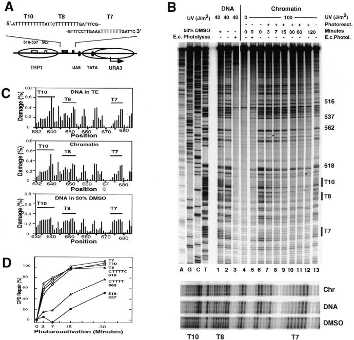
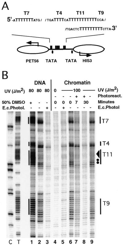
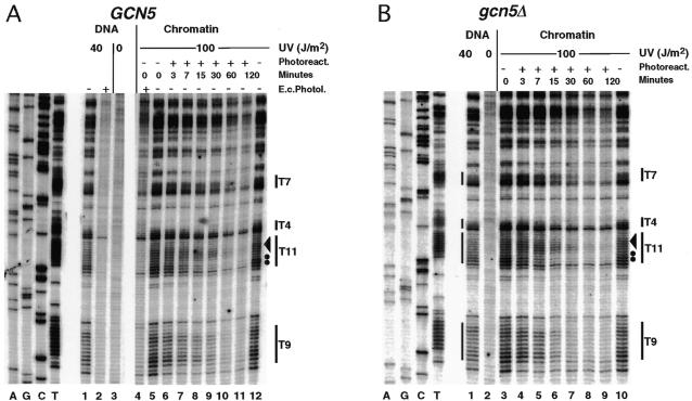
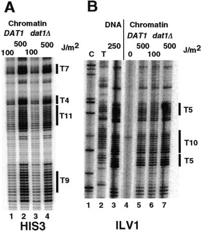
Poly(dA.dT) sequences (T-tracts) are abundant genomic DNA elements with unusual properties in vitro and an established role in transcriptional regulation of yeast genes. In vitro T-tracts are rigid, contribute to DNA bending, affect assembly in nucleosomes and generate a characteristic pattern of CPDs (cyclobutane pyrimidine dimers) upon irradiation with UV light (UV photofootprint). In eukaryotic cells, where DNA is packaged in chromatin, the DNA structure of T-tracts is unknown. Here we have used in vivo UV photofootprinting and DNA repair by photolyase to investigate the structure and accessibility of T-tracts in yeast promoters (HIS3, URA3 and ILV1). The same characteristic photofootprints were obtained in yeast and in naked DNA, demonstrating that the unusual T-tract structure exists in living cells. Rapid repair of CPDs in the T-tracts demonstrates that these T-tracts were not folded in nucleosomes. Moreover, neither datin, a T-tract binding protein, nor Gcn5p, a histone acetyltransferase involved in nucleosome remodelling, showed an influence on the structure and accessibility of T-tracts. The data support a contribution of this unusual DNA structure to transcriptional regulation.


Figures

Figure 1
Figure 1
UV photofootprints and repair analysis of T-tracts in the URA3 promoter of strain JMY1 [rad1Δ; YRpTRURAP (URA3, ARS1)]. (A) Schematic drawing of the chromatin structure and DNA elements in the top strand including nucleosome positions (ovals; overlapping positions at the TATA box) and T-tracts (T10, T8 and T7) (29,30). (B) Primer extension products showing damage formation and repair. UV damage generated by UV light (preferentially CPDs and 6–4PPs) in naked DNA: lane 1, in 50% DMSO; lane 2, in TE buffer; lane 6, in living cells (chromatin). Lanes 7–12, UV photoproducts after photoreactivation for 3–120 min in vivo. Lanes 3 and 5, non-CPDs (preferentially 6–4PPs) revealed by treatment with E.coli photolyase. Lane 4, DNA of non-irradiated cells. A, G, C and T, sequencing lanes. The T-tract regions of lanes 1, 2 and 6 are enlarged in the bottom panel. (C) Quantitative analysis. The graphs show the percentage of total DNA damage generated at individual dipyrimidine sequences. (D) DNA repair curves. Repair of CPDs is plotted versus incubation time (min) in the presence of photoreactivating light. The pyrimidine clusters 562 and 516–537 are located in the flanking nucleosome.
Figure 2
Figure 2
UV photofootprints and repair analysis of T-tracts in the extrachromosomal HIS3 promoter of strain FTY117 [rad1Δ; YRpCS1 (HIS3 TRP1 ARS1)]. (A) Schematic drawing of chromatin structure and DNA elements (top strand; according to 22). (B) Primer extension products. For space reasons, only the lower part of the gel is shown.
Figure 3
Figure 3
UV photofootprints and repair analysis in the genomic HIS3 promoter in the presence and absence of Gcn5p. (A) Primer extension products of strain GSY2 (rad1Δ GCN5). (B) Primer extension products of strain GSY5 (rad1Δ gcn5Δ). Dots and the arrowhead indicate fast and more slowly repaired sites within T11, respectively.
Figure 4
Figure 4
T-tract structure is independent of datin. (A) Primer extension analysis of UV photoproducts in the HIS3 promoter of strains BSY1 (DAT1, YRpCS1) and BSY2 (dat1Δ, YRpCS1). (B) Primer extension analysis of UV photoproducts in the ILV1 promoter of the same strains.

Similar articles

Cited by

References

    1. Nelson H.C., Finch,J.T., Luisi,B.F. and Klug,A. (1987) Nature, 330, 221–226. - PubMed
    1. DiGabriele A.D., Sanderson,M.R. and Steitz,T.A. (1989) Proc. Natl Acad. Sci. USA, 86, 1816–1820. - PMC - PubMed
    1. Leroy J.L., Charretier,E., Kochoyan,M. and Gueron,M. (1988) Biochemistry, 27, 8894–8898. - PubMed
    1. Alexeev D.G., Lipanov,A.A. and Skuratovskii,I. (1987) Nature, 325, 821–823. - PubMed
    1. Peck L.J. and Wang,J.C. (1981) Nature, 292, 375–378. - PubMed

Publication types

MeSH terms