

LEUNIG, a putative transcriptional corepressor that regulates AGAMOUS expression during flower development
- PMID: 11058164
- PMCID: PMC18862
- DOI: 10.1073/pnas.230352397
LEUNIG, a putative transcriptional corepressor that regulates AGAMOUS expression during flower development
Abstract
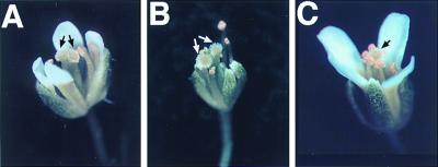
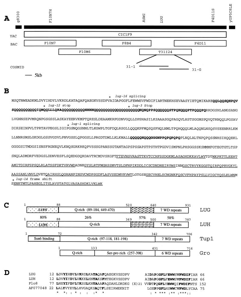
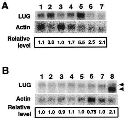
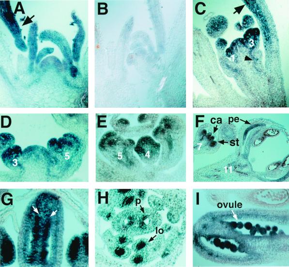
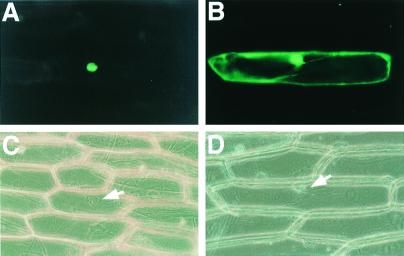
Regulation of homeotic gene expression is critical for proper developmental patterns in both animals and plants. LEUNIG is a key regulator of the Arabidopsis floral homeotic gene AGAMOUS. Mutations in LEUNIG cause ectopic AGAMOUS mRNA expression in the outer two whorls of a flower, leading to homeotic transformations of floral organ identity as well as loss of floral organs. We isolated the LEUNIG gene by using a map-based approach and showed that LEUNIG encodes a glutamine-rich protein with seven WD repeats and is similar in motif structure to a class of functionally related transcriptional corepressors including Tup1 from yeast and Groucho from Drosophila. The nuclear localization of LEUNIG-GFP is consistent with a role of LEUNIG as a transcriptional regulator. The detection of LEUNIG mRNA in all floral whorls at the time of their inception suggests that the restricted activity of LEUNIG in the outer two floral whorls must depend on interactions with other spatially restricted factors or on posttranslational regulation. Our finding suggests that both animals and plants use similar repressor proteins to regulate critical developmental processes.
Figures
Similar articles
-
Repression of AGAMOUS by BELLRINGER in floral and inflorescence meristems.Plant Cell. 2004 Jun;16(6):1478-89. doi: 10.1105/tpc.021147. Epub 2004 May 21. Plant Cell. 2004. PMID: 15155890 Free PMC article.
-
SEUSS, a member of a novel family of plant regulatory proteins, represses floral homeotic gene expression with LEUNIG.Development. 2002 Jan;129(1):253-63. doi: 10.1242/dev.129.1.253. Development. 2002. PMID: 11782418
-
LEUNIG regulates AGAMOUS expression in Arabidopsis flowers.Development. 1995 Apr;121(4):975-91. doi: 10.1242/dev.121.4.975. Development. 1995. PMID: 7743940
-
Transcriptional repression of target genes by LEUNIG and SEUSS, two interacting regulatory proteins for Arabidopsis flower development.Proc Natl Acad Sci U S A. 2004 Aug 3;101(31):11494-9. doi: 10.1073/pnas.0403055101. Epub 2004 Jul 26. Proc Natl Acad Sci U S A. 2004. PMID: 15277686 Free PMC article.
-
The N-Terminus of the Floral Arabidopsis TGA Transcription Factor PERIANTHIA Mediates Redox-Sensitive DNA-Binding.PLoS One. 2016 Apr 29;11(4):e0153810. doi: 10.1371/journal.pone.0153810. eCollection 2016. PLoS One. 2016. PMID: 27128442 Free PMC article.
Cited by
-
Repression of AGAMOUS by BELLRINGER in floral and inflorescence meristems.Plant Cell. 2004 Jun;16(6):1478-89. doi: 10.1105/tpc.021147. Epub 2004 May 21. Plant Cell. 2004. PMID: 15155890 Free PMC article.
-
SEUSS and AINTEGUMENTA mediate patterning and ovule initiation during gynoecium medial domain development.Plant Physiol. 2008 Mar;146(3):1165-81. doi: 10.1104/pp.107.114751. Epub 2008 Jan 9. Plant Physiol. 2008. PMID: 18184731 Free PMC article.
-
Transcriptional changes in litchi (Litchi chinensis Sonn.) inflorescences treated with uniconazole.PLoS One. 2017 Apr 18;12(4):e0176053. doi: 10.1371/journal.pone.0176053. eCollection 2017. PLoS One. 2017. PMID: 28419137 Free PMC article.
-
Fruit development in Arabidopsis.Arabidopsis Book. 2006;4:e0075. doi: 10.1199/tab.0075. Epub 2006 Feb 22. Arabidopsis Book. 2006. PMID: 22303227 Free PMC article. No abstract available.
-
SEUSS and LEUNIG regulate cell proliferation, vascular development and organ polarity in Arabidopsis petals.Planta. 2006 Sep;224(4):801-11. doi: 10.1007/s00425-006-0264-6. Epub 2006 Apr 20. Planta. 2006. PMID: 16625397
References
-
- Weigel D, Meyerowitz E M. Cell. 1994;78:203–209. - PubMed
-
- Yanofsky M F, Ma H, Bowman J L, Drews G N, Feldmann K A, Meyerowitz E M. Nature (London) 1990;346:35–39. - PubMed
-
- Bowman J L, Smyth D R, Meyerowitz E M. Development (Cambridge, UK) 1991;112:1–20. - PubMed
-
- Drews G N, Bowman J L, Meyerowitz E M. Cell. 1991;65:991–1002. - PubMed
-
- Liu Z, Meyerowitz E M. Development (Cambridge, UK) 1995;121:975–991. - PubMed
Publication types
MeSH terms
Substances
Associated data
- Actions
LinkOut - more resources
Full Text Sources
Other Literature Sources
Molecular Biology Databases
