Comparative Study
. 2000 Nov 1;19(21):5650-60.
doi: 10.1093/emboj/19.21.5650.

Crystal structure of the human alpha-thrombin-haemadin complex: an exosite II-binding inhibitor

Affiliations
Comparative Study

Crystal structure of the human alpha-thrombin-haemadin complex: an exosite II-binding inhibitor

J L Richardson et al. EMBO J. .

Abstract

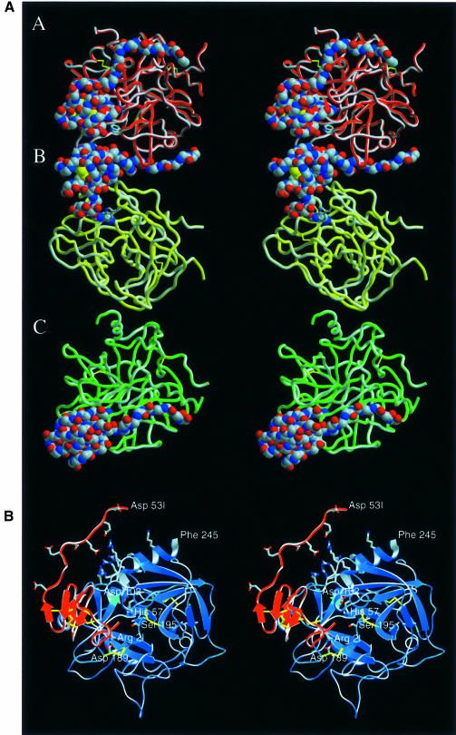
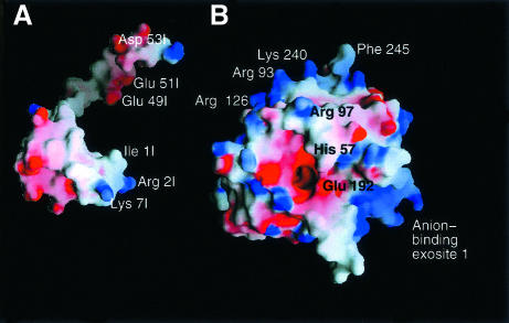
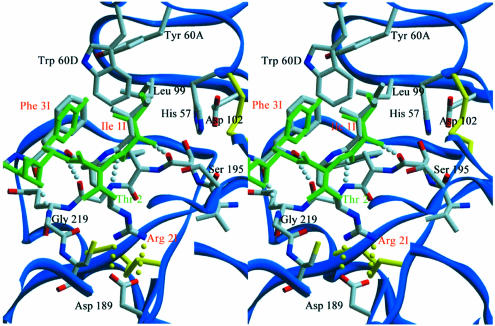
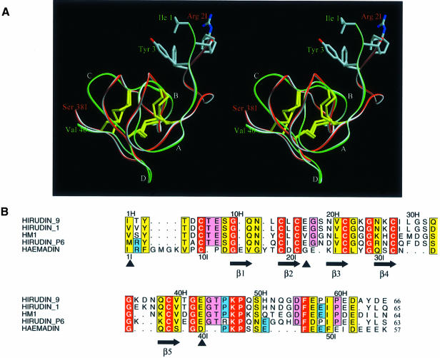
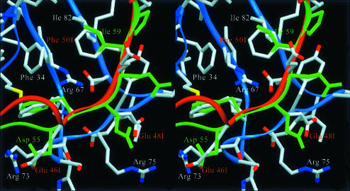
The serine proteinase alpha-thrombin plays a pivotal role in the regulation of blood fluidity, and therefore constitutes a primary target in the treatment of various haemostatic disorders. Haemadin is a slow tight- binding thrombin inhibitor from the land-living leech Haemadipsa sylvestris. Here we present the 3.1 A crystal structure of the human alpha-thrombin- haemadin complex. The N-terminal segment of haemadin binds to the active site of thrombin, forming a parallel beta-strand with residues Ser214-Gly216 of the proteinase. This mode of binding is similar to that observed in another leech-derived inhibitor, hirudin. In contrast to hirudin, however, the markedly acidic C-terminal peptide of haemadin does not bind the fibrinogen-recognition exosite, but interacts with the heparin-binding exosite of thrombin. Thus, haemadin binds to thrombin according to a novel mechanism, despite an overall structural similarity with hirudin. Haemadin inhibits both free and thrombomodulin-bound alpha-thrombin, but not intermediate activation forms such as meizothrombin. This specific anticoagulant ability of haemadin makes it an ideal candidate for an antithrombotic agent, as well as a starting point for the design of novel antithrombotics.


Figures

None
Fig. 1. Crystal structure of the human thrombin–haemadin complex. (A) Structure of the crystallographic trimer present in the asymmetric unit. Monomers are labelled A, B and C. Thrombin molecules are shown as red, yellow and green ribbons; the Cα traces of the three inhibitors are presented as colour-coded van der Waals spheres (red, oxygen; blue, nitrogen; grey, carbon). (B) Stereo diagram of complex molecule A. The protease is shown in its ‘standard orientation’ (Bode et al., 1992), i.e. with the active-site cleft facing the viewer and substrates running from left to right. Side chains of the catalytic triad residues are shown explicitly, as well as the side chains of the interacting residues Asp189 (thrombin) and Arg2I (haemadin) (colour coded as in Figure 1A). Also shown (unlabelled) are the side chains of the basic residues of the heparin-binding site (thrombin), as well as the side chains of the acidic residues of haemadin’s C-terminal tail. This figure and Figures 3, 5A, 7A and 8 were prepared with SETOR (Evans, 1993).
None
Fig. 2. Primary and secondary structure of haemadin. Acidic, basic and cysteine residues are represented by red, blue and yellow circles, respectively. Main chain–main chain hydrogen bonds formed in at least two of the three independent molecules forming the asymmetric unit are indicated with dotted lines. Disulfide bridges are shown with yellow lines. Residues involved in interactions with thrombin are represented by grey circles.
None
Fig. 3. Ribbon diagram of haemadin’s structure, highlighting elements of secondary structure and the disulfide-rich core. Side chains of residues involved in major interactions with thrombin are shown explicitly (colour coded as in Figure 1).
None
Fig. 4. Space-filling models of human α-thrombin and haemadin, showing the surface potential of the two molecules. Positive charges are displayed in blue, negative charges in red, with darkest blue and red colours corresponding to electrostatic potentials beyond –10 and +10 kT/e, respectively. The plot was prepared with GRASP (Nicholls et al., 1993). The thrombin component (B) is shown in the standard orientation, while haemadin (A) is rotated along the y-axis to present the thrombin binding surface to the viewer. Some of the residues of the interacting interfaces are labelled.
None
Fig. 5. Close-up stereoview of the active-site cleft of haemadin-bound human α-thrombin. Side chains of selected residues are depicted colour coded. Intermolecular hydrogen bonds are shown as white dotted lines for the parallel interaction of haemadin’s N-terminal loop with thrombin residues Ser214–Gly216 or yellow dotted lines for contacts made inside the S1 pocket. Some residues of thrombin and the inhibitors along with all water molecules are omitted for the sake of simplicity. The three N-terminal amino acid residues of hirudin (green) are shown for comparison.
None
Fig. 6. Binding studies in solution. (A) Formation of the ternary complex haemadin–thrombin–triabin, as followed using non-denaturant PAGE. Lane 1, human α-thrombin (5 µg); lane 2, haemadin (10 µg); lane 3, triabin (20 µg); lane 4, thrombin–haemadin complex (10 µg); lanes 5–7, 10 µg of thrombin–haemadin complex incubated with increasing amounts of triabin (1:1, 1:1.5 and 1:2 equivalents); lane 9, human α-thrombin–triabin complex (10 µg). (B) Haemadin binds to thrombomodulin-bound thrombin, but not to meizothrombin. Lane 1, 10 µg of human thrombin–TME456; lane 2: 10 µg of the complex incubated with 1 µg haemadin; lane 3, 10 µg of meizothrombin; lane 4, 10 µg meizothrombin incubated with 1.5 µg haemadin; lane 5, 20 µg triabin; lane 6, human thrombin–triabin complex (10 µg).
None
Fig. 7. (A) Stereoview of the main chain of haemadin (red, residues Ile1I–Ser38I) and hirudin (green, residues Ile1H–Val40H) after optimal least-squares fit; only the side chains of the first three residues of both molecules are shown explicitly. Note the different location of the N-terminal segments, indicating divergent arrangements of the compact domains relative to thrombin (compare Figure 5). (B) Structure-based alignment of the amino acid sequences of haemadin and of four representative hirudin variants. Nomenclature follows the work of Steiner et al. (1992). Residues with particularly close homologies are boxed in yellow, identities in red. Residues conserved in hirudin but not haemadin are shadowed pink; those common to haemadin and some hirudin variants are shadowed blue. Numbers refer to the sequences of hirudin (above) and haemadin (below the alignment). The secondary structure of haemadin is also given. The intron–exon boundaries (full arrows) are those determined for Hirudinaria manillensis (Scacheri et al., 1993). The aligned sequences were formatted using the program ALSCRIPT (Barton, 1993).
None
Fig. 8. Close-up stereoview comparing the interactions of the C-terminal tails of haemadin (red) and hirudin (green) with the fibrinogen-recognition exosite of a neighbouring thrombin molecule (blue) (see text for details). Side chains of interacting thrombin/inhibitor residues are labelled explicitly. Notice the close agreement between the phenyl moieties of Phe47I and Phe56H; also the side chain pairs Phe50I–Ile59H and Glu48I–Glu57H occupy similar positions.

Similar articles

Cited by

References

    1. Arocas V., Zingali,R.B., Guillin,M.-C., Bon,C. and Jandrot-Perrus,M. (1996) Bothrojaracin: a potent two-site-directed thrombin inhibitor. Biochemistry, 35, 9083–9089. - PubMed
    1. Barton G.J. (1993) ALSCRIPT: a tool to format multiple sequence alignments. Protein Eng., 6, 37–40. - PubMed
    1. Betz A., Hofsteenge,J. and Stone,S.R. (1992) Interaction of the N-terminal region of hirudin with the active-site cleft of thrombin. Biochemistry, 31, 4557–4562. - PubMed
    1. Bode C., Nordt,T.K. and Runge,M.S. (1994) Thrombolytic therapy in acute myocardial infarction—selected recent developments. Ann. Hematol., 69, S35–40. - PubMed
    1. Bode W. and Huber,R. (1992) Natural protein proteinase inhibitors and their interaction with proteinases. Eur. J. Biochem., 204, 433–451. - PubMed

Publication types

MeSH terms