. 2000 Nov 1;19(21):5661-71.
doi: 10.1093/emboj/19.21.5661.

Unprecedented proximal binding of nitric oxide to heme: implications for guanylate cyclase

Affiliations

Unprecedented proximal binding of nitric oxide to heme: implications for guanylate cyclase

D M Lawson et al. EMBO J. .

Abstract

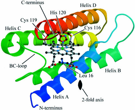
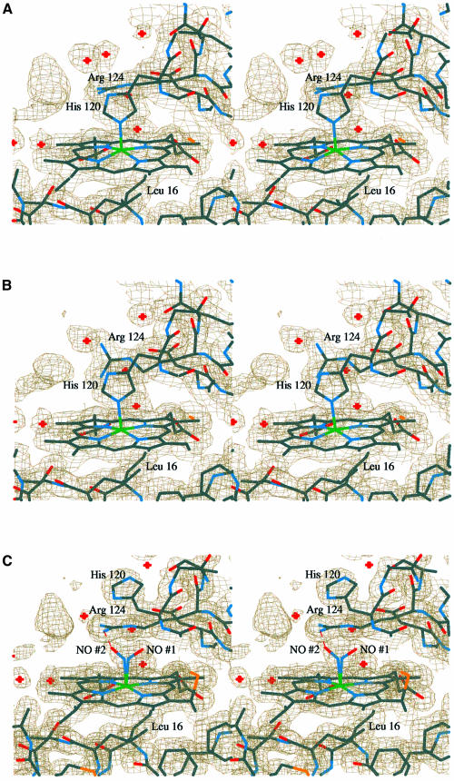
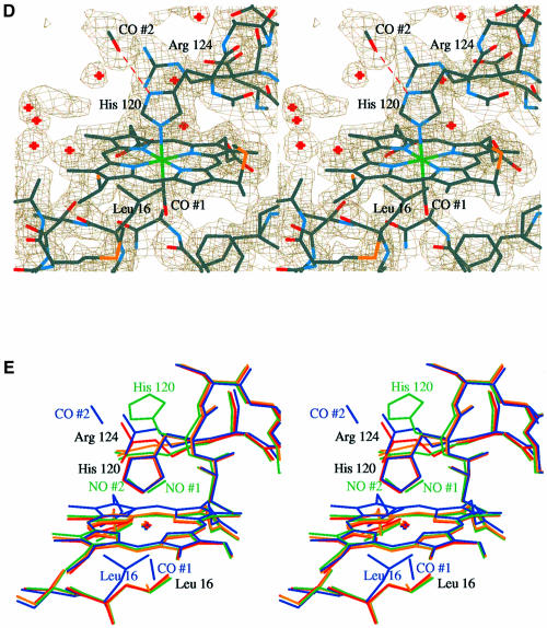
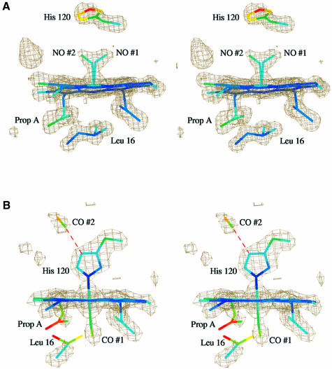
Microbial cytochromes c' contain a 5-coordinate His-ligated heme that forms stable adducts with nitric oxide (NO) and carbon monoxide (CO), but not with dioxygen. We report the 1.95 and 1.35 A resolution crystal structures of the CO- and NO-bound forms of the reduced protein from Alcaligenes xylosoxidans. NO disrupts the His-Fe bond and binds in a novel mode to the proximal face of the heme, giving a 5-coordinate species. In contrast, CO binds 6-coordinate on the distal side. A second CO molecule, not bound to the heme, is located in the proximal pocket. Since the unusual spectroscopic properties of cytochromes c' are shared by soluble guanylate cyclase (sGC), our findings have potential implications for the activation of sGC induced by the binding of NO or CO to the heme domain.


Figures

None
Fig. 1. Ribbon representation of a single subunit of the reduced Axcyt c′ structure showing the position of the heme. Also depicted are the side chains of the proximal His, Leu16 and the two Cys residues that form thioether bridges to the heme. The Leu blocks access to the vacant sixth coordination site in the distal pocket. The location of the crystallographic 2-fold axis is indicated, which is perpendicular to the plane of the paper. A 180° rotation of this subunit about the 2-fold axis generates the second subunit of the functional dimer. The structure is colored with respect to sequence number, starting with blue at the N-terminus and finishing with red at the C-terminus. This figure was produced using MOLSCRIPT (Kraulis, 1991) and Raster3D (Merritt and Bacon, 1997).
None
Fig. 2. Stereo diagrams of the final 2mFobs – dFcalc electron density maps contoured at 1σ for the (A) oxidized, (B) reduced, (C) NO-bound and (D) CO-bound Axcyt c′ structures. The atoms are colored as follows: Fe, green; S, yellow; O, red; N, blue; C, gray. The red crosses indicate water molecules. In this view, both the left-hand and rear edges of the heme are accessible to solvent, as is evident from the presence of water molecules in these regions. In the interests of clarity, the thioether bridges to the heme group, which would be in the foreground, have been omitted. In the oxidized and NO-bound structures in particular, there is a large unassigned region of electron density within hydrogen bonding distance of the side chain of Arg124. Attempts to model buffer and cryoprotectant components here were not convincing. However, a reasonable fit was obtained with a sulfate anion (present in the precipitant), but this gave high temperature factors after refinement. (E) A least squares superposition of all four structures based on all main chain atoms. They are colored as follows: oxidized, yellow; reduced, red; NO-bound, green; CO-bound, blue. In the interests of clarity, all bonds to the heme Fe have been omitted. All images were produced using the programs O and OPLOT (Jones et al., 1991).
None
Fig. 3. Omit difference maps for the (A) NO- and (B) CO-bound Axcyt c′ structures contoured at 3.5σ. All the displayed atoms were omitted from the refinements (see Materials and methods). However, Arg124 was retained in the refinement, in order to remove distracting density, in particular from the NO ligand. The view is chosen to emphasize the flattening of the heme plane upon binding of CO. The atoms are colored according to their temperature factors, which all lie roughly in the range 10–40 Å2, where dark blue indicates a low value increasing through light blue, dark green, light green, yellow and orange, to red, which indicates a high value. Note that the side chain of His120 in the NO-bound structure and the side chain of Leu16 and the propionate group A in the CO-bound structure are less well defined in the electron density and have relatively high thermal parameters. See main text for a further explanation.

Similar articles

Cited by

References

    1. Addison A.W. and Stephanos,J.J. (1986) Nitrosyliron(III) hemoglobin: autoreduction and spectroscopy. Biochemistry, 25, 4104–4113. - PubMed
    1. Ambler R.P. (1973) The amino acid sequence of cytochrome c′ from Alcaligenes sp. NCIMB 11015. Biochem. J., 135, 751–758. - PMC - PubMed
    1. Brucker E.A., Olson,J.S., Ikeda-Saito,M. and Phillips,G.N.,Jr (1998) Nitric oxide myoglobin: crystal structure and analysis of ligand geometry. Proteins, 30, 352–356. - PubMed
    1. Brünger A.T. (1992) X-PLOR: Version 3.1. A System for X-ray Crystallography and NMR. Yale University Press, New Haven, CT.
    1. Brünger A.T. (1993) Assessment of phase accuracy by cross validation—the free R-value—methods and applications. Acta Crystallogr. D, 49, 24–36. - PubMed

Publication types

MeSH terms