. 2000 Nov 1;19(21):5711-9.
doi: 10.1093/emboj/19.21.5711.

The Rab6-binding kinesin, Rab6-KIFL, is required for cytokinesis

Affiliations

The Rab6-binding kinesin, Rab6-KIFL, is required for cytokinesis

E Hill et al. EMBO J. .

Abstract

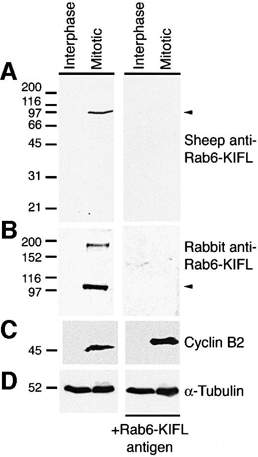
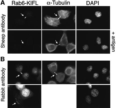
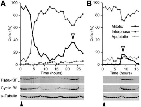
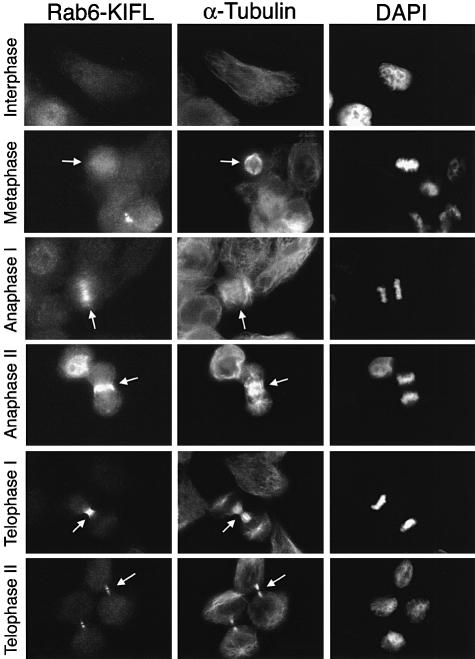
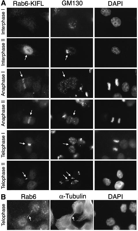
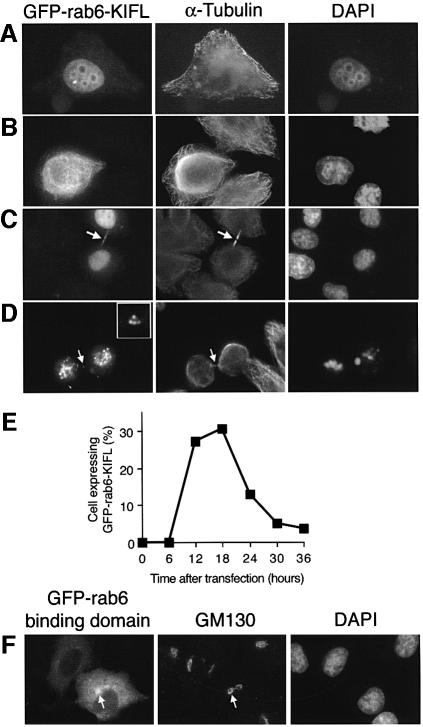
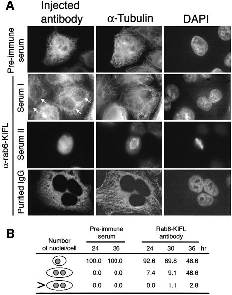
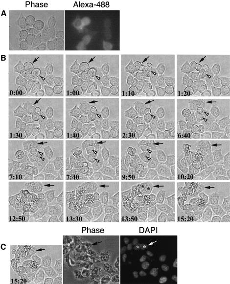
The Rab6-binding kinesin, Rab6-KIFL, was identified in a two-hybrid screen for proteins that interact with Rab6, a small GTPase involved in membrane traffic through the Golgi apparatus. We find that Rab6-KIFL accumulates in mitotic cells where it localizes to the midzone of the spindle during anaphase, and to the cleavage furrow and midbody during telophase. Overexpression of Rab6-KIFL causes a cell division defect resulting in cell death. Microinjection of antibodies to Rab6-KIFL results in the cells becoming binucleate after one cell cycle, and time-lapse microscopy reveals that this is due to a defect in cleavage furrow formation and thus cytokinesis. These data show that endogenous Rab6-KIFL functions in cell division during cleavage furrow formation and cytokinesis, in addition to its previously described role in membrane traffic.


Figures

None
Fig. 1. Characterization of Rab6-KIFL antibodies. (A) Protein extracts, 100 µg, from either interphase or nocodazole-arrested mitotic HeLa cells, >90% in mitosis, were analysed by western blotting with an affinity-purified sheep polyclonal antibody raised against Rab6-KIFL. For control blots the antibody was pre-incubated with 50 µg of the recombinant protein antigen. (B) HeLa cell extracts were western blotted as above with the published rabbit polyclonal antibody to Rab6-KIFL again with a control in which the antibody was pre-incubated with its antigen. (C and D) The same samples were also probed for cyclin B2 as a marker for mitotic arrest, and α-tubulin as a loading control, respectively.
None
Fig. 2. Rab6-KIFL localizes to the midbody in dividing cells. (A) HeLa cells were stained with the affinity-purified sheep polyclonal antibody to Rab6-KIFL, a monoclonal antibody to α-tubulin and DAPI to reveal the DNA. Control coverslips were stained as above, except that the antibody dilution was pre-incubated with 25 µg of the Rab6-KIFL antigen for 1 h on ice. (B) HeLa cells were stained as above except that the published rabbit antibody to Rab6-KIFL was used in place of the sheep antibody. Arrows denote areas of particular interest.
None
Fig. 3. Rab6-KIFL levels oscillate in a cell cycle controlled manner. (A) HeLa cells were arrested with nocodazole for 20 h, then released into fresh drug-free medium. Samples were collected every hour as shown, and the number of mitotic, interphase and apoptotic cells counted. Cell extracts were analysed by western blotting with antibodies to Rab6-KIFL, cyclin B2 and α-tubulin as a loading control. (B) HeLa cells were arrested with 2 mM hydroxyurea for 18 h, then released into fresh drug-free medium. The cells were analysed as before. Solid arrowheads indicate the time of drug washout, and open arrowheads the peak of mitotic cells.
None
Fig. 4. Rab6-KIFL localizes to the spindle midzone during anaphase and cleavage furrow during cytokinesis. HeLa cells were stained with the affinity-purified sheep antibody to Rab6-KIFL, a monoclonal antibody to α-tubulin, while the DNA was stained with DAPI. Representative images of cells at the indicated stages of the cell cycle are shown. Arrows denote areas of particular interest.
None
Fig. 5. Discrete localization of Rab6-KIFL and the Golgi apparatus throughout the cell cycle. (A) HeLa cells fixed with methanol were stained with the affinity-purified sheep antibody to Rab6-KIFL, a rabbit polyclonal antibody to the Golgi marker GM130, while the DNA was stained with DAPI. Representative images of cells at the indicated stages of the cell cycle are shown. (B) HeLa cells fixed with paraformaldehyde were stained with antibodies to Rab6 and α-tubulin; the DNA was stained with DAPI. A representative image of cells at the indicated stage of the cell cycle is shown. Arrows denote areas of particular interest.
None
Fig. 6. Overexpression of Rab6-KIFL interferes with normal cell division. (AD) HeLa cells were transfected with an expression plasmid for GFP-tagged Rab6-KIFL. Cells were fixed in methanol after 18 h and processed for immunofluorescence with antibodies to α-tubulin. Representative images demonstrating the various effects of Rab6-KIFL overexpression are shown. (E) Quantitation of the number of cells expressing GFP–Rab6-KIFL as a function of time after transfection. (F) HeLa cells were transfected with an expression plasmid for the GFP-tagged Rab6-binding domain of Rab6-KIFL. Cells were fixed in methanol after 18 h and processed for immunofluorescence with antibody to GM130. A representative image demonstrating the effect of overexpressing the Rab6-binding domain construct is shown.
None
Fig. 7. Effects of antibody microinjection on cell division. (A) HeLa cells grown on coverslips were microinjected as described in Materials and methods with pre-immune serum, the Rab6-KIFL antiserum or purified sheep immunoglobulin (IgG) to Rab6-KIFL. Representative images of microinjected cells are shown for each condition, 36 h post-injection. (B) The number of normal, binucleate or multinucleate microinjected cells was counted at the times indicated, after microinjection of either the pre-immune serum or the affinity-purified antibody to Rab6-KIFL. The numbers shown are from a typical experiment in which 200 cells were microinjected for each condition. In each case similar results were obtained for three independent experiments performed with all the conditions shown.
None
Fig. 8. Time-lapse imaging of cells microinjected with antibodies to Rab6-KIFL. (A) HeLa cells grown on coverslips were microinjected as described in Materials and methods with the Rab6-KIFL antibody and purified donkey IgG conjugated to Alexa-488. Cells were then allowed to recover for 6 h before imaging. A phase contrast, and corresponding Alexa-488 fluorescence image are shown. (B) Time-lapse imaging of the cells shown in (A) above; time after commencement of imaging is indicated in hours and minutes. The filled arrow indicates a microinjected cell in mitosis that has started to divide. The open arrowhead indicates a non-injected cell in mitosis that divides normally. (C) After 15 h 20 min of imaging the cells were fixed and the DNA stained with DAPI. The same cells are shown as in (B) above. A movie corresponding to this figure is available as Supplementary data at The EMBO Journal Online.

Similar articles

Cited by

References

    1. Adams R.R., Tavares,A.A., Salzberg,A., Bellen,H.J. and Glover,D.M. (1998) pavarotti encodes a kinesin-like protein required to organize the central spindle and contractile ring for cytokinesis. Genes Dev., 12, 1483–1494. - PMC - PubMed
    1. Allan V.J. and Schroer,T.A. (1999) Membrane motors. Curr. Opin. Cell Biol., 11, 476–482. - PubMed
    1. Barr F.A. (1999) A novel Rab6-interacting domain defines a family of Golgi-targeted coiled-coil proteins. Curr. Biol., 9, 381–384. - PubMed
    1. Boman A.L., Kuai,J., Zhu,X., Chen,J., Kuriyama,R. and Kahn,R.A. (1999) Arf proteins bind to mitotic kinesin-like protein 1 (MKLP1) in a GTP-dependent fashion. Cell Motil. Cytoskel., 44, 119–132. - PubMed
    1. Burgess R.W., Deitcher,D.L. and Schwarz,T.L. (1997) The synaptic protein syntaxin1 is required for cellularization of the Drosophila embryo. J. Cell Biol., 138, 861–875. - PMC - PubMed

Publication types

LinkOut - more resources