. 2000 Nov 1;19(21):5793-800.
doi: 10.1093/emboj/19.21.5793.

Non-enzymatic triggering of the ceramide signalling cascade by solar UVA radiation

Affiliations

Non-enzymatic triggering of the ceramide signalling cascade by solar UVA radiation

S Grether-Beck et al. EMBO J. .

Abstract

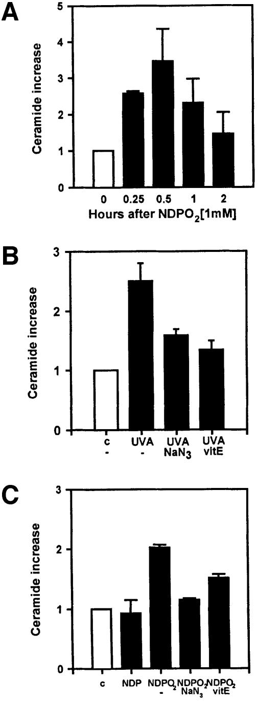
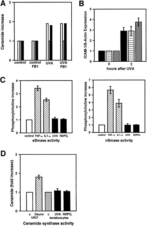
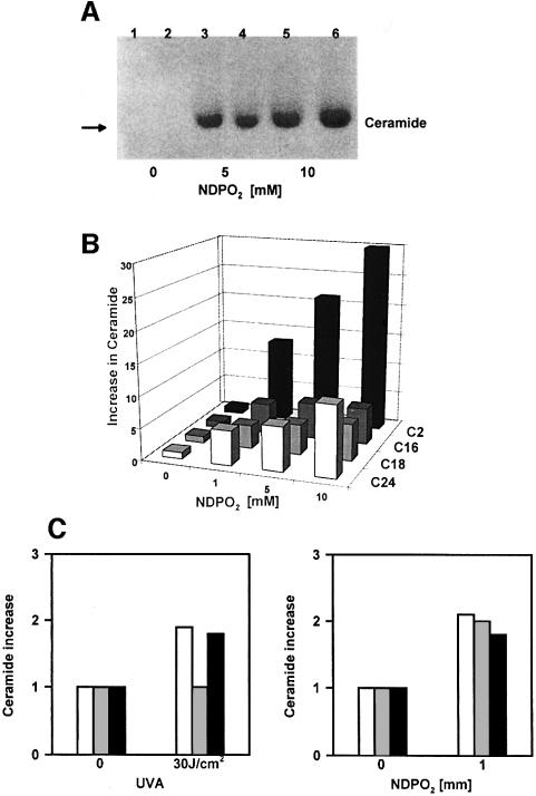
Ceramide is a key component of intracellular stress responses. Evidence is provided for a novel mechanism of ceramide formation that mediates solar ultraviolet (UV) A radiation-induced expression of the intercellular adhesion molecule (ICAM)-1. Similarly to UVA radiation, ceramide stimulation of human keratinocytes induced ICAM-1 mRNA expression and activated the ICAM-1 promoter through transcription factor AP-2. Ceramide-activated AP-2 and ceramide-induced ICAM-1 reporter gene activation were abrogated through deletion of the AP-2 binding site. UVA radiation increased the level of ceramide in keratinocytes and inhibition of sphingomyelin synthesis prevented UVA radiation-induced ICAM-1 expression. Hitherto, two pathways have been identified for ceramide accumulation: hydrolysis from sphingomyelin through neutral and acid sphingomyelinases, and de novo synthesis by ceramide synthase. UVA radiation did not activate any of these enzymes. Ceramide generation in UVA-irradiated cells, however, was inhibited by singlet oxygen quenchers and mimicked in unirradiated cells by a singlet oxygen-generating system. In addition, UVA radiation and singlet oxygen both generated ceramide in protein-free, sphingomyelin-containing liposomes. This study indicates that singlet oxygen triggers a third, non-enzymatic mechanism of ceramide formation.


Figures

None
Fig. 1. Role of ceramide in UVA radiation-induced ICAM-1 expression in long-term cultured, normal human keratinocytes. (A) Time-dependent activation of transcription factor AP-2 after stimulation of cells with 10 µM C2-ceramide. Nuclear extracts were analysed by electrophoretic mobility shift assay using a radiolabelled AP-2 consensus site derived from the human ICAM-1 promoter. (B) Time-dependent ICAM-1 mRNA expression after stimulation of cells with 10 µM C2-ceramide (grey bar). ICAM-1 mRNA expression was assessed by differential RT–PCR based on the housekeeping gene β-actin. Unstimulated control (white bar) is set as 1. (C) C2-ceramide-induced reporter gene activity in transiently transfected human keratinocytes detected as relative specific luciferase activity (RLU/µg protein) (grey bars). Different deletion constructs based on the human ICAM-1 promoter linked to luciferase were assessed in triplicate. Unstimulated but transfected cells were set as 1 (white bar). (D) UVA-induced ceramide release was detected in human keratinocytes by sequential HPTLC in triplicate. Cells were sham irradiated (white bar) or irradiated with 30 J/cm2 UVA (black bars) and harvested at the indicated time points. Data were obtained as c.p.m./500 µg of protein (mean of three experiments) and are given as fold increase (control was set as 1). (E) Effect of l-cycloserine on UVA-induced ICAM-1 mRNA expression. UVA radiation-induced ICAM-1 mRNA expression was assessed by differential RT–PCR in cells that had been left untreated (black bar) or treated with 1 mM l-cycloserine (striped bar) for 3 days prior to, during and after UVA irradiation, or were additionally stimulated with 10 µM C2-ceramide (grey bar).
None
Fig. 2. Singlet oxygen-induced release of ceramide in long-term cultured, normal human keratinocytes. (A) Time-dependent release of ceramide after a 30 min exposure of cells to 1 mM NDPO2. Unstimulated control cells are set as 1 (white bar). Measurements were made in triplicate. Effect of sodium azide (50 mM, added during stimulation) and vitamin E (25 µM, added as α-tocopheryl succinate 24 h prior to stimulation) on UVA radiation- (B) and NDPO2 (1 mM)-induced (C) release of ceramide (black bars). Ceramide release was assessed 30 min after stimulation. Unstimulated controls are set as 1 (white bar).
None
Fig. 3. The role of ceramide synthase and sphingomyelinase activation in ceramide formation by long-term cultured, normal human keratinocytes. (A) UVA-induced ceramide (C24, white bar; C18, grey bar; C16, black bar) release was detected after lipid extraction by HPLC/mass spectroscopy in cells that had been incubated with 25 µM fumonisin B1 during and after irradiation (30 J/cm2 UVA). (B) Expression of ICAM-1 mRNA was assessed in cells that had been left untreated (black bar) or incubated with 25 µM fumonisin B1 without (striped bar) or with 10 µM C2-ceramide (grey bar) during and 30 min after irradiation (30 J/cm2 UVA). (C) Activation of acid and neutral sphingomyelinase was measured in cells 30 min after stimulation with cytokines [hatched bars; 1000 U/ml recombinant human (rh) TNF-α, 1000 U/ml rh IL-1α from R&D Systems], UVA radiation (30 J/cm2) or 1 mM NDPO2 (black bars). Unstimulated controls were set as 1 (white bars). (D) Activation of ceramide synthase was measured in vitro in microsomal membranes of U937 cells that had been left untreated (open bar) or stimulated with 1 µM daunorubicin for 24 h (hatched bar), and in keratinocytes that had been left untreated (grey bar) or stimulated with either 30 J/cm2 UVA or NDPO2 (black bars), and harvested after 30 min.
None
Fig. 4. Singlet oxygen generates ceramide in sphingomyelin-containing liposomes. (A) HPTLC of NDPO2-induced ceramide formation in liposomes. Liposomes were treated with 0 (lanes 1 and 2), 5 (lanes 3 and 4) and 10 mM NDPO2 (lanes 5 and 6), and chloroform/methanol lipid extracts prepared immediately thereafter and analysed. Iodine vapour was used to stain ceramides. (B) Analysis of NDPO2-induced ceramide formation in liposomes by HPLC/mass spectroscopy. Liposomes were treated for 30 min with increasing doses (0–10 mM) of NDPO2 and lipid extracts prepared immediately thereafter. (C) Analysis of ceramide in long-term cultured human keratinocytes by HPLC/mass spectroscopy. Lipid extracts were prepared 30 min after stimulation of cells with 30 J/cm2 UVA or 1 mM NDPO2. C24, white bars; C18, grey bars; C16, black bars.
None
Fig. 5. Absolute amounts of ceramide and sphingomyelin in liposomes and human keratinocytes. (A) The absolute amounts of C2- (open circle), C16- (black circle), C18- (dark grey circle) and C24- (grey circle) ceramides were determined in picomoles in sphingomyelin-containing liposomes that had been treated with increasing concentrations of NDPO2 (0, 1, 5 and 10 mM) as described in Materials and methods. Data are given as the generation of ceramide (pmol)/sphingomyelin (µmol) versus increasing concentrations of NDPO2 (mM). (B) Time-dependent increase of absolute amounts of ceramide (grey bar) and sphingomyelin (black bar) in human keratinocytes. Cells were either left untreated or stimulated with 1 mM NDPO2, and subsequently the absolute amounts of ceramide and sphingomyelin determined as described in Materials and methods. All measurements were made in triplicate. Data are given in the form of a histogram of amount of lipid (pmol)/500 µg of protein versus time (h) after stimulation with 1 mM NDPO2.

References

    1. Ahrens C. et al. (1997) Photocarcinogenesis and inhibition of intercellular adhesion molecule 1 expression in cells of DNA-repair defective individuals. Proc. Natl Acad. Sci. USA, 94, 6837–6841. - PMC - PubMed
    1. Anderson V.C. and Thompson,D.H. (1992) Triggered release of hydrophilic agents from plasmalogen liposomes using visible light or acid. Biochim. Biophys. Acta, 1109, 33–42. - PubMed
    1. Ballou L.R., Laulederkind,S.J.F., Rosloniec,E.F. and Raghow,R. (1996) Ceramide signalling and the immune response. Biochim. Biophys. Acta, 1301, 273–287. - PubMed
    1. Bender K., Blattner,C., Knebel,A., Iordanov,M., Herrlich,P. and Rahmsdorf,H.J. (1997) UV-induced signal transduction. J. Photochem. Photobiol. B, 37, 1–17. - PubMed
    1. Berneburg M., Grether-Beck,S., Kürten,V., Ruzicka,T., Briviba,K., Sies,H. and Krutmann,J. (1999) Singlet oxygen mediates the UVA-induced generation of the photoaging-associated mitochondrial common deletion. J. Biol. Chem., 274, 15345–15349. - PubMed

Publication types

MeSH terms