. 2000 Nov 1;19(21):5895-904.
doi: 10.1093/emboj/19.21.5895.

Human pre-mRNA cleavage factor II(m) contains homologs of yeast proteins and bridges two other cleavage factors

Affiliations

Human pre-mRNA cleavage factor II(m) contains homologs of yeast proteins and bridges two other cleavage factors

H de Vries et al. EMBO J. .

Abstract

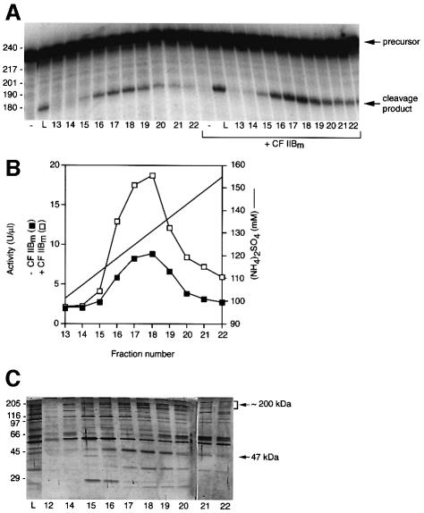
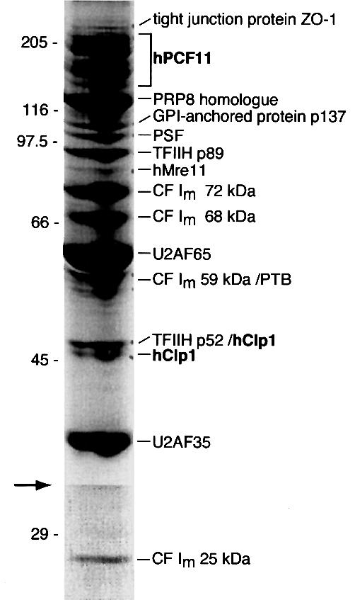
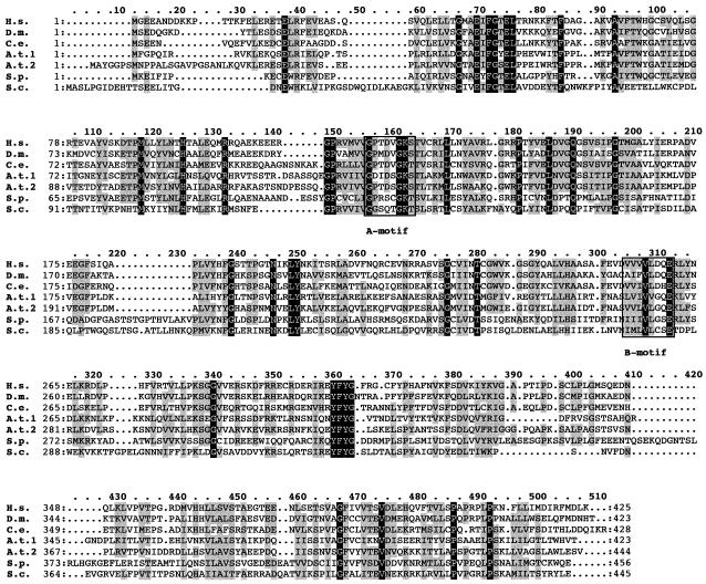
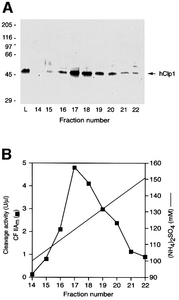
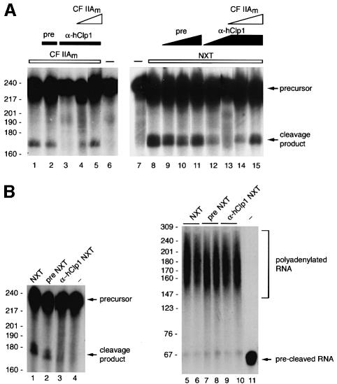
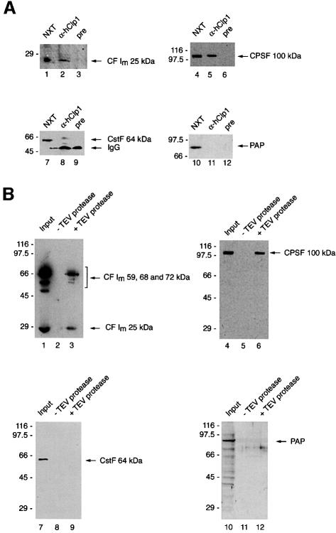
Six different protein factors are required in vitro for 3' end formation of mammalian pre-mRNAs by endonucleolytic cleavage and polyadenylation. Five of the factors have been purified and most of their components cloned, but cleavage factor II(m) (CF II(m)) remained uncharacterized. We have purified CF II(m) from HeLa cell nuclear extract by several chromatographic steps. During purification, CF II(m) activity separated into two components, one essential (CF IIA(m)) and one stimulatory (CF IIB(m)) for the cleavage reaction. CF IIA(m) fractions contain the human homologs of two yeast 3' end processing factors, Pcf11p and Clp1p, as well as cleavage factor I(m) (CF I(m)) and several splicing and transcription factors. We report the cloning of hClp1 and show that it is a genuine subunit of CF IIA(m). Antibodies directed against hClp1 deplete cleavage activity, but not polyadenylation activity from HeLa cell nuclear extract. hClp1 interacts with CF I(m) and the cleavage and polyadenylation specificity factor CPSF, suggesting that it bridges these two 3' end processing factors within the cleavage complex.


Figures

None
Fig. 1. Purification scheme of CF IIm. The chromatographic columns used to separate CF IIAm and CF IIBm and to purify CF IIAm are shown schematically. The concentrations and types of salts for the preparation of HeLa cell NXT and during the fractionation are indicated below each step.
None
Fig. 2. Purification of CF IIAm by Mono Q column chromatography. (A) CF IIAm activity profile over the Mono Q column with and without the addition of CF IIBm. Cleavage assays were carried out as described in Materials and methods for 85 min at 30°C with SV40 late pre-mRNA as substrate and 8 µl of the fractions indicated at the bottom. Samples to which 2 µl of CF IIBm were added are marked with a bracket. Samples were analyzed on a denaturing 6% (w/v) polyacryl amide gel. Sizes of standards in nucleotides are indicated on the left. –, SV40 late pre-mRNA incubated with all protein factors except CF IIAm; L, 4 µl of the load of the Mono Q column. (B) Quantitation of the assay shown in (A). The Mono Q fractions were assayed in the presence (open squares) or absence (filled squares) of 2 µl of CF IIBm. Activities (U/µl) were determined as described (Rüegsegger et al., 1996). (C) Silver stain of a 10% SDS–polyacrylamide gel resolving proteins from the Mono Q fractions. Aliquots of 2 µl of the fractions indicated at the bottom were loaded. The molecular masses of the size standards in kDa are indicated on the left. Polypeptides co-eluting with CF IIAm activity are indicated with an arrowhead and a bracket. L, the load of the Mono Q column.
None
Fig. 3. Identification of the polypeptides in CF IIAm. A peak fraction of the final Mono Q column was separated on a 10% SDS–polyacryl amide gel and the gel was stained with colloidal Coomassie. The lower part of the image (indicated with an arrow) was processed differently to compensate for the weaker staining of low molecular mass polypeptides. The molecular masses of the size standards in kDa are indicated on the left. All polypeptides were identified by mass spectrometry and are labeled accordingly on the right. Polypeptides highlighted in bold co-elute precisely with CF IIAm activity on Mono Q. The accession numbers are: tight junction protein ZO-1, SwissProt (sp): Q07157; hPcf11, DDBJ/EMBL/GenBank (gb): AB020631; PRP8 homolog, sp: O60231; GPI-anchored protein p137, sp: Q14444; PSF, sp: P23246; TFIIH p89, sp: P19447; hMre11, sp: P49959; CF Im 68 kDa, gb: X67337; U2AF65, sp: P26368; PTB, sp: P26599; TFIIH p52, TREMBL (tr): Q92759; hClp1, tr: Q92989; U2AF35, sp: Q01081; CF Im 25 kDa, tr: O43809. No database accession numbers have been assigned yet to the CF Im 59 and 72 kDa proteins.
None
Fig. 4. hClp1 is evolutionarily conserved and has an ATP/GTP-binding motif. The alignment of H.sapiens (tr: Q92989), D.melanogaster (tr: Q9V6Q1), C.elegans (sp: P52874), A.thaliana 1 (gb: AB010077), A.thaliana 2 (tr: QSR06), S.pombe (tr: Q10299) and S.cerevisiae (tr: Q08685) Clp1p sequences was generated with clustalx. The black and gray boxes indicate identical and similar residues, respectively. The conserved Walker A motif with the consensus sequence -A/G-X-X-X-X-G-K-S/T- and the B motif are indicated.
None
Fig. 5. hClp1 co-elutes with CF IIAm activity from a Mono Q column. In contrast to the profile shown in Figure 2B, the load of this column was a pool from a phenyl Superose column and the Mono S and Ni-NTA steps were omitted. (A) Aliquots of 10 µl of the column fractions indicated at the bottom were separated on a 10% SDS–polyacrylamide gel and blotted onto a nitrocellulose membrane, which was detected with α-hClp1. The molecular masses of the size standards in kDa are indicated on the left. (B) Cleavage activity profile of the Mono Q column. Assays were carried out as described in Materials and methods for 85 min at 30°C with 2 µl of the fractions indicated at the bottom and L3 pre-mRNA as substrate. Samples were analyzed on a denaturing 6% (w/v) polyacrylamide gel and the CF IIAm activity (filled squares) was determined as described in Materials and methods.
None
Fig. 6. Antibodies directed against hClp1 deplete CF IIAm activity from partially purified CF IIAm and dialyzed HeLa cell NXT. Cleavage assays were carried out as described in Materials and methods for 90 min at 30°C with SV40 late pre-mRNA as substrate. Samples were analyzed on a denaturing 6% (w/v) polyacrylamide gel. Sizes of standards in nucleotides are indicated on the left. (A) Immunodepletion experiments with partially purified CF IIAm (lanes 1–5) and dialyzed NXT (lanes 8–15). Lanes 1–5, SV40 late pre-mRNA was incubated in the presence of CF Im, CstF, CPSF, PAP and the CF IIAm fraction indicated. Lane 1, 4 µl of CF IIAm (phenyl Superose column fraction, input of the immunodepletion experiments); lane 2, 4 µl of CF IIAm depleted with pre-immune serum; lane 3, 4 µl of CF IIAm depleted with α-hClp1; lane 4, 2 µl of CF IIAm depleted with α-hClp1 plus 2 µl of CF IIAm (phenyl Superose column fraction, same as input); lane 5, 2 µl of CF IIAm depleted with α-hClp1 plus 4 µl of CF IIAm (phenyl Superose column fraction, same as input); lanes 6 and 7, SV40 late pre-mRNA incubated in the absence of protein factors; lane 8, 3 µl of NXT (input of the immunodepletion experiments); lanes 9–11, 3 µl of NXT depleted with increasing amounts of pre-immune serum (5, 10 and 15 µl of beads, respectively); lanes 12 and 13, 3 µl of NXT depleted with increasing amounts of α-hClp1 (5 and 10 µl of beads, respectively); lanes 14 and 15, 3 µl of NXT depleted with α-hClp1 (10 µl of beads) to which increasing amounts of CF IIAm were added (2 and 4 µl of a phenyl Superose fraction). (B) hClp1 is not essential for polyadenylation in vitro. Extracts depleted with pre-immune serum or α-hClp1 were tested for cleavage (lanes 1–3) and polyadenylation (lanes 5–10). Polyadenylation assays were carried out as described in Materials and methods for 30 min at 30°C with L3 pre-cleaved mRNA as substrate. All samples were analyzed on denaturing 6% (w/v) polyacrylamide gels. Lanes 1, 5 and 6, 2 µl of dialyzed NXT (input of the immunodepletion experiments); lanes 2, 7 and 8, 2 µl of NXT depleted with pre-immune serum; lanes 3, 9 and 10, 2 µl of NXT depleted with α-hClp1; lane 4, SV40 late pre-mRNA; lane 11, L3 pre-cleaved mRNA.
None
Fig. 7. CF Im and CPSF interact with hClp1. (A) Western blot analysis of immunoprecipitations with α-hClp1 and pre-immune serum, respectively. Proteins were separated on 10% SDS–polyacrylamide gels, blotted onto nitrocellulose membranes, and the blots were probed with α-CF Im25 kDa, α-CPSF100 kDa, α-CstF64 kDa or α-PAP, respectively. Lane 1, 1 µl of NXT (10% of the input of the immunoprecipitations); lane 2, 40% of the immunoprecipitate (α-hClp1); lane 3, 40% of the immunoprecipitate (pre); lanes 4, 7 and 10, 3 µl of NXT (3% of the input of the immunoprecipitations); lanes 5, 8 and 11, 50% of the immunoprecipitate (α-hClp1); lanes 6, 9 and 12, 50% of the immuno precipitate (pre). (B) Binding experiments with GST–TEV-tagged hClp1 were carried out as described in Materials and methods. The samples were separated on a 10% SDS–polyacrylamide gel and blotted onto a nitrocellulose membrane, which was probed with α-CF Im68 kDa and α-CF Im25 kDa, α-CPSF100 kDa, α-CstF64 kDa or α-PAP, respectively. Lane 1, 5 µl of CF Im (30% of input); lane 2, 14 µl of the mock digest (28%); lane 3, 14 µl of the digest with TEV protease (28%); lane 4, 5 µl of CPSF (30% of input); lane 7, 5 µl of CstF (10% of input); lane 10, 3 µl of PAP (10% of input); lanes 5, 8 and 11, 14 µl of the mock digest (20%); lanes 6, 9 and 12, 14 µl of the digest with TEV protease (20%). The molecular masses of the size standards in kDa are indicated on the left.

Similar articles

Cited by

References

    1. Amrani N., Minet,M., Wyers,F., Dufour,M.E., Aggerbeck,L.P. and Lacroute,F. (1997) PCF11 encodes a third protein component of yeast cleavage and polyadenylation factor I. Mol. Cell. Biol., 17, 1102–1109. - PMC - PubMed
    1. Barabino S.M. and Keller,W. (1999) Last but not least: regulated poly(A) tail formation. Cell, 99, 9–11. - PubMed
    1. Barabino S.M., Hübner,W., Jenny,A., Minvielle-Sebastia,L. and Keller,W. (1997) The 30-kD subunit of mammalian cleavage and polyadenylation specificity factor and its yeast homolog are RNA-binding zinc finger proteins. Genes Dev., 11, 1703–1716. - PubMed
    1. Bienroth S., Wahle,E., Suter-Crazzolara,C. and Keller,W. (1991) Purification of the cleavage and polyadenylation factor involved in the 3′-end processing of messenger RNA precursors. J. Biol. Chem., 266, 19768–19776. - PubMed
    1. Fountoulakis M. and Langen,H. (1997) Identification of proteins by matrix-assisted laser desorption ionization-mass spectrometry following in-gel digestion in low-salt, nonvolatile buffer and simplified peptide recovery. Anal. Biochem., 250, 153–156. - PubMed

Publication types

MeSH terms