. 2000 Nov 1;19(21):5905-15.
doi: 10.1093/emboj/19.21.5905.

Progression of a loop-loop complex to a four-way junction is crucial for the activity of a regulatory antisense RNA

Affiliations

Progression of a loop-loop complex to a four-way junction is crucial for the activity of a regulatory antisense RNA

F A Kolb et al. EMBO J. .

Abstract

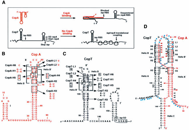
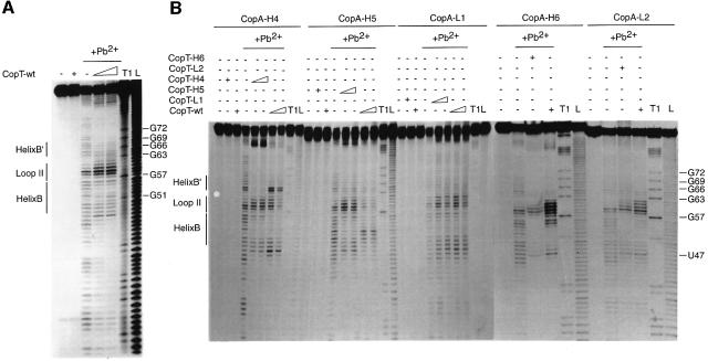
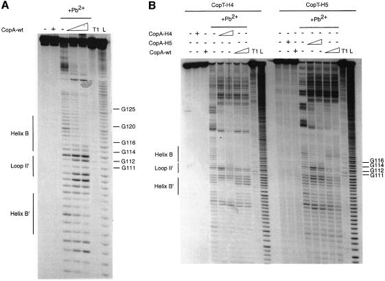
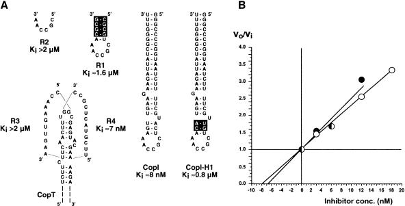
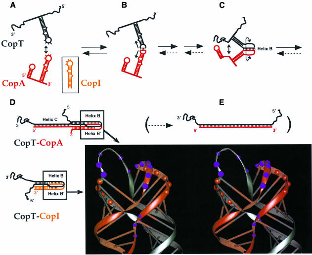
The antisense RNA, CopA, regulates the replication frequency of plasmid R1 through inhibition of RepA translation by rapid and specific binding to its target RNA (CopT). The stable CopA-CopT complex is characterized by a four-way junction structure and a side-by-side alignment of two long intramolecular helices. The significance of this structure for binding in vitro and control in vivo was tested by mutations in both CopA and CopT. High rates of stable complex formation in vitro and efficient inhibition in vivo required initial loop-loop complexes to be rapidly converted to extended interactions. These interactions involve asymmetric helix progression and melting of the upper stems of both RNAs to promote the formation of two intermolecular helices. Data presented here delineate the boundaries of these helices and emphasize the need for unimpeded helix propagation. This process is directional, i.e. one of the two intermolecular helices (B) must form first to allow formation of the other (B'). A binding pathway, characterized by a hierarchy of intermediates leading to an irreversible and inhibitory RNA-RNA complex, is proposed.


Figures

None
Fig. 1. Schematic model for antisense RNA control of RepA synthesis (A) and secondary structures of the antisense RNA CopA (B), its target site CopT (C) and the stable CopA–CopT complex (D). (A) Binding of CopA prevents ribosome binding at the tap ribosomal binding site (RBS), and the presence of a stem–loop structure sequesters the repA RBS so that translation of RepA is inhibited. In the absence of CopA binding, translation of tap permits ribosome entry at the repA loading site (translational coupling). SD, Shine–Dalgarno sequence. (B–D) The mutated nucleotides (H1–H6, L1 and L2) are boxed. The secondary structure model for the stable CopA–CopT complex (D) was derived from chemical and enzymatic probing (Kolb et al., 2000). RNase V1 cleavages in CopA and CopT that occur in the CopA–CopT complex are shown by arrows: increased cleavages are shown by red arrows, unchanged cleavages are shown by black arrows; Pb2+-induced cleavages in the CopA–CopT complex are shown by blue circles.
None
Fig. 2. Pb2+-induced hydrolysis of homologous or heterologous CopA–CopT complexes using end-labelled CopA species. Hydrolysis was performed on 5′-end-labelled wild-type CopA (A) or CopA-H4, CopA-H5, CopA-L1, CopA-H6 and CopA-L2 (B), alone (–) or in the presence of an excess of wild-type or mutant CopT (+). Complex formation was performed at 37°C for 15 min in TMN buffer. Lanes T1 and L, RNases T1 and alkaline ladders, respectively.
None
Fig. 3. Pb2+-induced hydrolysis of homologous or heterologous CopA–CopT complexes using end-labelled CopT species. Hydrolysis was performed on 5′-end-labelled wild-type CopT (A) or CopT-H4 and CopT-H5 (B), alone (–) or in the presence of an excess of wild-type or mutant CopA (+). Lanes T1 and L, RNases T1 and alkaline ladders, respectively.
None
Fig. 4. Competitive inhibition between CopA and different RNA variants for the formation of the stable CopA–CopT complex. (A) The inhibitory RNAs used in this study and their corresponding Ki values are shown. Experimental conditions are described in detail in Materials and methods. Nucleotides that are not complementary to CopT are boxed. (B) Determination of the Ki values for CopI and RNA R4. The ratio vo/vi was calculated from the inhibition experiments using different concentrations of CopI (open circles) or of RNA R4 (closed circles) at time intervals between 1 and 6 min at 37°C, and are plotted against the inhibitor concentrations.
None
Fig. 5. The binding pathway of CopA–CopT. (AE) refer to steps in the pathway as explained in the Discussion. The parentheses indicate that full duplex formation is slow and biologically irrelevant (step E). Dotted arrows denote very slow reactions. The structure model of the four-helix junction derived from computer modelling by Kolb et al. (2000) is shown. Red and purple circles represent the RNase V1 and Pb2+-induced cleavages.

Similar articles

Cited by

References

    1. Asano K. and Mizobuchi,K. (2000) Structural analysis of late intermediate complex formed between plasmid ColIb-P9 Inc RNA and its target RNA. J. Biol. Chem., 275, 1269–1274. - PubMed
    1. Asano K., Kato,A., Morikawi,H., Hama,C., Shiba,K. and Mizobuchi,K. (1991) Positive and negative regulations of plasmid ColIb-P9 repZ gene expression at the translational level. J. Biol. Chem., 266, 3774–3781. - PubMed
    1. Asano K., Niimi,T., Yokoyama,S. and Mizobuchi,K. (1998) Structural basis for binding of the plasmid ColIb-P9 antisense Inc RNA to its target RNA with the 5′-rUUGGCG-3′ motif in the loop sequence. J. Biol. Chem., 273, 11826–11838. - PubMed
    1. Ashraf S.S., Ansari,G., Guenther,R., Sochacka,E., Malkiewicz,A. and Agris,P.F. (1999) The uridine in ‘U-turn’: Contributions to tRNA–ribosomal binding. RNA, 5, 503–511. - PMC - PubMed
    1. Berzal-Herranz A., Wagner,E.G.H. and Diaz-Orejas,R. (1991) Control of replication of plasmid R1: the intergenic region between copA and repA modulates the level of expression of repA. Mol. Microbiol., 5, 97–108. - PubMed

Publication types