Comparative Study
. 2000 Nov;38(11):4096-101.
doi: 10.1128/JCM.38.11.4096-4101.2000.

Detection of influenza A viruses from different species by PCR amplification of conserved sequences in the matrix gene

Affiliations
Comparative Study

Detection of influenza A viruses from different species by PCR amplification of conserved sequences in the matrix gene

R A Fouchier et al. J Clin Microbiol. 2000 Nov.

Abstract

The recently raised awareness of the threat of a new influenza pandemic has stimulated interest in the detection of influenza A viruses in human as well as animal secretions. Virus isolation alone is unsatisfactory for this purpose because of its inherent limited sensitivity and the lack of host cells that are universally permissive to all influenza A viruses. Previously described PCR methods are more sensitive but are targeted predominantly at virus strains currently circulating in humans, since the sequences of the primer sets display considerable numbers of mismatches to the sequences of animal influenza A viruses. Therefore, a new set of primers, based on highly conserved regions of the matrix gene, was designed for single-tube reverse transcription-PCR for the detection of influenza A viruses from multiple species. This PCR proved to be fully reactive with a panel of 25 genetically diverse virus isolates that were obtained from birds, humans, pigs, horses, and seals and that included all known subtypes of influenza A virus. It was not reactive with the 11 other RNA viruses tested. Comparative tests with throat swab samples from humans and fecal and cloacal swab samples from birds confirmed that the new PCR is faster and up to 100-fold more sensitive than classical virus isolation procedures.


Figures

FIG. 1
FIG. 1
Entropy plots of oligonucleotide-annealing sites in human and animal influenza A virus sequences available from the influenza virus sequence database. The sequences recognized by oligonucleotides Mx1, Fam1, NS486C, Mx2, Fam2, NS637R, M52C, M253R, and M93C were compared to all available influenza A virus sequences (n = 189, 189, 234, 203, 204, 249, 175, 215, and 189, respectively), and their heterogeneities are displayed in panels A through I, respectively. Oligonucleotide positions are given in the 5′ to 3′ direction, with position 1 being the extreme 5′ nucleotide. Asterisks indicate primer positions with degeneracy in the designed oligonucleotides. Oligonucleotides M52C, M253R, and M93C were designed in the present study.
FIG. 2
FIG. 2
PCR analysis of the influenza A viruses, listed in Table 1, which originated from different hosts and geographical locations. RNA was isolated from influenza A viruses grown in embryonated chicken eggs, followed by PCR analysis and agarose gel electrophoresis (top panels) or dot blot analysis (bottom panels). Lanes 1 to 25, see Table 1; lane 26, negative control.
FIG. 3
FIG. 3
Sensitivity of detection of influenza A virus RNA by PCR. RNA was isolated from 0.2 ml of 10-fold serial dilutions of influenza virus A/Netherlands/18/94 (107 TCID50s/ml) and was used for PCR analysis followed by agarose gel electrophoresis and ethidium bromide staining (top panel) or dot blot analysis (bottom panel). Lane 1, negative control; lanes 2 to 9, dilution series representing the equivalent of 2 × 10⁵ to 0.02 TCID50s per sample. Samples containing less than 0.02 TCID50 were negative by PCR and dot blot analysis (data not shown).
FIG. 4
FIG. 4
Specificity of detection of influenza A virus RNA by PCR. RNA was isolated from virus stocks and was used for PCR analysis and subsequent agarose gel electrophoresis (top panel) or dot blot hybridization (bottom panel). Lanes: 1, HRSV; 2, PIV-1; 3, PIV-2; 4, PIV-3; 5, PIV-4; 6, Sendai virus; 7, SV5; 8, NDV; 9, mumps virus; 10, measles virus; 11, influenza B virus; 12, influenza A virus.
FIG. 5
FIG. 5
PCR-based detection of influenza A virus in 39 human throat swab samples. Throat swab samples that were tested previously for the presence of influenza A virus by classical screening methods (7) were randomized and tested blindly by PCR. RNA was isolated from 0.2 ml of a throat swab sample and was used for PCR and dot blot analysis. Lanes 1, 4, 7, 8, 13, 16, 18, 23, 24, 30, 34, 35, and 38, influenza virus-negative samples; lanes 2, 5, 9, 10, 12, 14, 15, 20, 21, 22, 25, 29, and 31, influenza B virus-positive samples; lane 40, 10 TCID50s of influenza virus A/Netherlands/18/94 as a positive control; lanes 3, 6, 11, 17, 19, 26, 27, 28, 32, 33, 36, 37, and 39, influenza A virus-positive samples in which virus titers determined in MDCK cells were 105.75, 0, 103.5, 102.25, 100.75, 104.25, 100.75, 103.75, 104.25, 105.25, 104.5, 105.75, and 103.5 TCID50s/ml respectively.
FIG. 6
FIG. 6
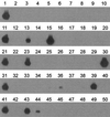
PCR-based detection of influenza A virus in a representative set of avian cloacal swab and dropping samples. RNA was isolated from 0.2 ml of 38 pooled samples, each consisting of five individual bird samples, and was used for PCR and Southern blot analysis. Lanes 1, 11, 21, 31, and 41, positive controls representing 10 TCID50s of influenza virus A/Netherlands/18/94; lanes 7, 14, 20, 27, 34, 40, and 47, negative controls; lanes 2 to 5, duck cloacal swab samples; lanes 6, 8 to 10, 12, 13, 15 to 19, 22 to 26, and 28 to 30, goose dropping samples; lanes 32, 33, 35 to 39, 42 to 46, and 48 to 50, goose cloacal swab samples. Each of the pools represented in lanes 13, 15, 23, 30, 36, 39, 43, and 44 was found to contain a single positive individual bird sample. Virus was isolated in embryonated chicken eggs from samples represented in lanes 13, 15, 23, 30, 39, and 43 but not from those represented in lanes 35, 36, and 44.

Comment in

References

    1. Atmar R L, Baxter B D, Dominguez E A, Taber L H. Comparison of reverse transcription-PCR with tissue culture and other rapid diagnostic assays for detection of type A influenza virus. J Clin Microbiol. 1996;34:2604–2606. - PMC - PubMed
    1. Brown T. Analysis of DNA sequences by blotting and hybridization. In: Ausubel F M, Brent R, Kingston R E, Moore D D, Seidman J G, Smith J A, Struhl K, editors. Current protocols in molecular biology, suppl. 45. New York, N.Y: John Wiley & Sons, Inc.; 2000. pp. 2.9.1–2.9.15.
    1. Cherian T, Bobo L, Steinhoff M C, Karron R A, Yolken R H. Use of PCR-enzyme immunoassay for identification of influenza A virus matrix RNA in clinical samples negative for cultivable virus. J Clin Microbiol. 1994;32:623–628. - PMC - PubMed
    1. Claas E C, Osterhaus A D. New clues to the emergence of flu pandemics. Nat Med. 1998;4:1122–1123. - PubMed
    1. Claas E C, Osterhaus A D, van Beek R, De Jong J C, Rimmelzwaan G F, Senne D A, Krauss S, Shortridge K F, Webster R G. Human influenza A H5N1 virus related to a highly pathogenic avian influenza virus. Lancet. 1998;351:472–477. - PubMed

Publication types