. 2000 Oct;11(2):59-76.
doi: 10.1002/1097-0193(200010)11:2<59::aid-hbm10>3.0.co;2-5.

Sources on the anterior and posterior banks of the central sulcus identified from magnetic somatosensory evoked responses using multistart spatio-temporal localization

Affiliations

Sources on the anterior and posterior banks of the central sulcus identified from magnetic somatosensory evoked responses using multistart spatio-temporal localization

M X Huang et al. Hum Brain Mapp. 2000 Oct.

Abstract

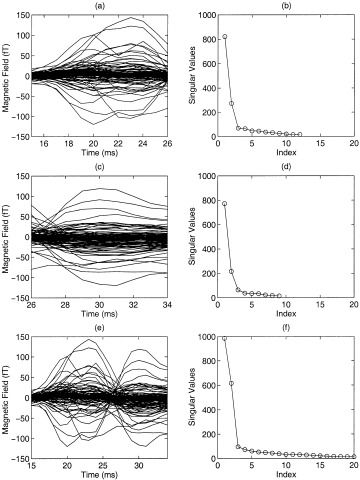
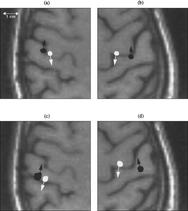
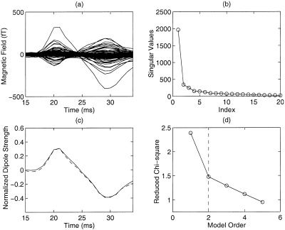
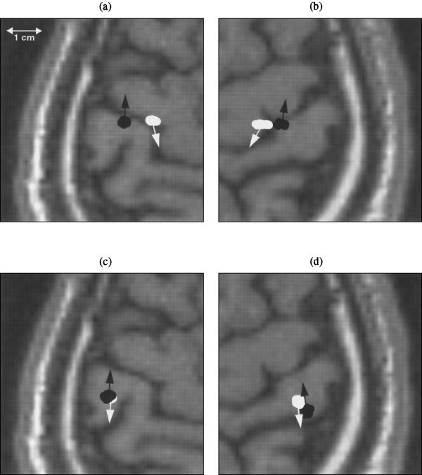
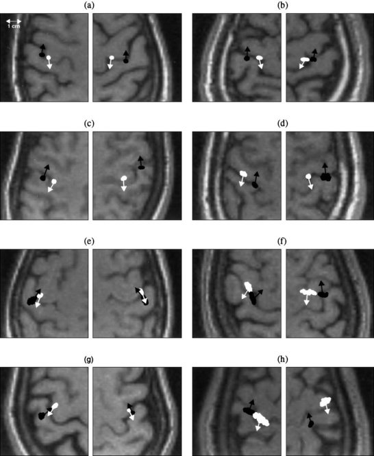
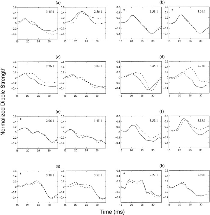
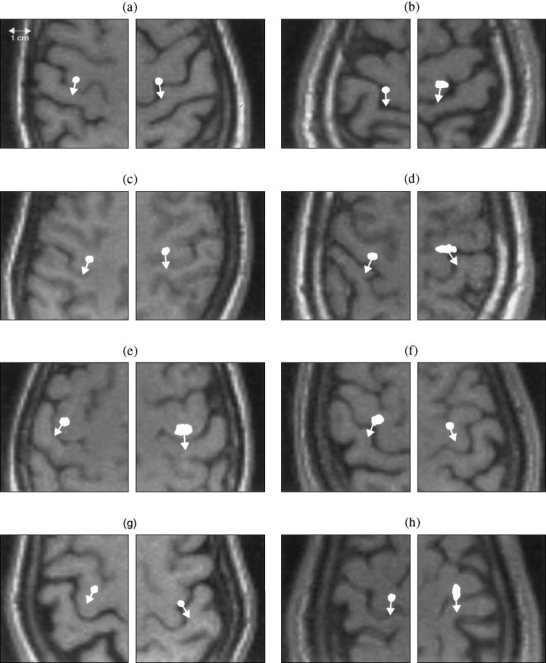
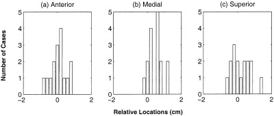
A Multi-Start Spatio-Temporal (MSST) multidipole localization algorithm was used to study sources on the anterior and posterior banks of the central sulcus localized from early somatosensory magnetoencephalography (MEG) responses. Electrical stimulation was applied to the right and left median nerves of 8 normal subjects. Two sources, one on the anterior and one on the posterior bank of the central sulcus, were localized from 16 data sets (8 subjects, 2 hemispheres). Compared with the more traditional practice of single-dipole fits to peak latencies, MSST provided more reliable source locations. The temporal dynamics of the anterior and posterior central sulcus sources, obtained using MSST, showed considerable temporal overlap. In some cases, the two sources appeared synchronous. On the other hand, in the traditional single-dipole peak-latency fit approach, there is no time course other than a focal dipole moment activated only at the selected peak latency. The same group of subjects also performed a motor task involving index-finger lifting; the anterior central sulcus source obtained from electrical median nerve stimulation localized to the same or similar region in the primary motor area identified from the finger-lift task. The physiological significance of the anterior central sulcus source is discussed. The findings suggest that one can test the integrity of cortical tissue in the region of primary motor cortex using electrical somatosensory stimulation.


Figures

Figure 1
Figure 1
A case of asynchronous anterior and posterior central sulcus sources. MEG waveforms and singular value plots of a subject's somatosensory responses for three intervals. (a)(b): Waveforms and singular value plot for the 15–26 ms poststimulus interval; (c)(d) for the 26–34 ms poststimulus interval; (e)(f) for the entire 15–34 ms interval.
Figure 2
Figure 2
Locations and orientations of asynchronous anterior and posterior central sulcus sources localized by Multi‐Start Spatio‐Temporal (MSST) 2‐dipole fit and a traditional peak‐latency single‐dipole fit for median nerve MEG responses. One hundred Monte‐Carlo analyses were performed to obtain the uncertainty of the locations, reflected by the size of the plotted locations. Radiological convention is adopted in the figures; left and right hemispheres are displayed on the right and left portions of the page, respectively. (a) Anterior and posterior central sulcus sources obtained by MSST for the left median nerve stimulation, indicated by white and black color, respectively. (b) Anterior and posterior central sulcus sources obtained by MSST for right median nerve stimulation. (c) Two sources obtained by traditional single‐dipole fit using the first and second major peak‐latencies for left median nerve response, indicated by black and white colors, respectively. Note that the source locations were almost an entire sulcus posterior to the MSST solutions. (d) Anterior and posterior central sulcus sources obtained by single dipole fit using the early two major peak‐latencies for right median nerve response.
Figure 3
Figure 3
A case of synchronous anterior and posterior central sulcus sources. (a) Waveforms of the subject for the 15–34 ms interval; (b) SVD only shows one signal‐related singular value; (c) Synchronous time courses of the anterior (dashed line) and posterior (solid line) central sulcal sources obtained by MSST using a 2‐dipole model; (d) Reduced Chi‐square plotted as a function of modeled dipoles. The vertical dashed line indicates that the data is appropriately modeled by 2 dipoles.
Figure 4
Figure 4
Locations and orientations of near synchronous anterior and posterior central sulcus sources localized by MSST and the traditional single dipole peak latency fit for one subject. (a) Anterior and posterior central sulcus sources obtained by MSST for the left median nerve response, indicated by white and black color, respectively. (b) Anterior and posterior central sulcus sources obtained by MSST for right median nerve response. (c) Two sources obtained by traditional single‐dipole fit using the first and second major peak‐latencies for left median nerve response, indicated by black and white colors, respectively. (d) Two sources obtained by single‐dipole fit using the first and second major peak‐latencies for right median nerve response.
Figure 5
Figure 5
Anterior and posterior central sulcus sources and their orientations localized by MSST for the left and right median nerve responses of 8 normal subjects. The anterior central sulcus sources are indicated by white color and posterior central sulcus sources by black color. One hundred Monte‐Carlo analyses were conducted for all the sources to obtain the uncertainty of the source locations.
Figure 6
Figure 6
Temporal dynamics of the anterior (dashed line) and posterior (solid lines) central sulcus sources obtained by using MSST on the left and right median nerve responses of eight normal subjects. For most of the 16 data sets (11), the posterior central sulcus sources preceded the anterior ones, but this temporal ordering was not evident in the remaining 5 cases (indicated by asterisks). All the responses show two peaks at about 20 ms and 30 ms with different polarities. The majority of them (except for the right and left responses for subjects (b)(d)(f) and the right response for subject (a)) also show a peak at about 25 ms with the same polarity as that of the 20 ms peak. The ratio of the time courses for the posterior central source vs. the anterior central source is shown in the upper‐right corner of each plot.
Figure 7
Figure 7
Primary motor sources and their orientations localized by MSST for the left and right index finger‐lifting responses of eight normal subjects. Again, 100 trial Monte‐Carlo analyses were conducted to obtain the uncertainty of the source locations. A comparison of these sources with the anterior central sulcus sources from median nerve responses (Fig. 5) reveal that either they are in the same regions, or the primary motor sources evoked by index‐finger lifts are slightly more medial.
Figure 8
Figure 8
Histogram plots of the relative locations of the primary motor sources from index‐finger lift task to the anterior central sulcus sources from median nerve responses. The sources were localized by MSST for eight normal subjects.
Figure 9
Figure 9
Fitting magnetic fields generated by two dipoles when fitted with a single dipole can cause observable mislocation. (a) The original 2‐dipole configuration; (b) A single dipole fit. The distance between this single dipole‐fitted location and the stronger dipole in (a) is 7.6 mm; (c) The magnetic field pattern generated by the 2‐dipole configuration; (d) Field pattern by the single dipole fit.

Similar articles

Cited by

References

    1. Achim A, Richer F, Saint‐Hilaire JM. (1991): Methodological consideration for the evaluation of spatio‐temporal source models. Electroenceph Clin Neurophysiol 79: 227–240. - PubMed
    1. Aine C, Huang M, Stephen J, Christner R. (2000): Multi‐start algorithms for MEG empirical data analysis reliably characterize locations and time‐courses of multiple sources. NeuroImage 12: 159–172. - PubMed
    1. Allison T, Wood CC, McCarthy G, Spencer DD. (1991a): Cortical somatosensory evoked potentials. II. Effects of excision of somatosensory or motor cortex in humans and monkeys. J Neurophysiol 66: 64–82. - PubMed
    1. Allison T, McCarthy G, Wood CC, Jones SJ. (1991b): Potentials evoked in human and monkey cerebral cortex by stimulation of the median nerve. Brain 114: 2465–2503. - PubMed
    1. Ahonen AI, Hämäläinen MS, Kajola MJ, Knuutila JET, Laine. (1992): A 122‐channel magnetometer covering the whole head. In: Dittmar A, Froment JC, eds): Proceedings of the Satellite Symposium on Neuroscience and Technology, 14th annual conference of IEEE Engineering in Medicine and Biology Society: IEEE Engineering and Medicine and Biology Society, pp. 16–20.

Publication types

LinkOut - more resources